
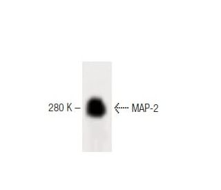


MAP-2 Antibody (18): sc-135979
- MAP-2 Antibody (18) is a mouse monoclonal IgG1, cited in 3 publications, provided at 50 µg/0.5 ml
- raised against amino acids 19-219 of MAP-2B of human origin
- recommended for detection of MAP-2B of mouse, rat and human origin by WB, IP and IF; not recommended for IP
- See MAP-2 (A-4): sc-74421 for MAP-2 antibody conjugates, including AC, HRP, FITC, PE, Alexa Fluor® 488, 594, 647, 680 and 790.
- At present, we have not yet completed the identification of the preferred secondary detection reagent(s) for MAP-2 Antibody (18). This work is in progress.

MAP-2 Antibody (18) is a mouse monoclonal IgG1 antibody that detects microtubule-associated protein 2 (MAP-2) in mouse, rat, and human samples through applications such as western blotting (WB), immunoprecipitation (IP), and immunofluorescence (IF). MAP-2 plays a crucial role in the stabilization and assembly of microtubules, which are essential components of the cytoskeletal network that provide structural support and shape to cells. MAP-2 is primarily located in neuronal cell bodies and dendrites, where MAP-2 is vital for maintaining the integrity of the neuronal cytoskeleton and facilitating intracellular transport. MAP-2 is particularly important in neuronal development and plasticity, as MAP-2 influences the morphology of dendritic spines and synaptic connections. Dysregulation of MAP-2 has been associated with various neurodegenerative diseases, highlighting MAP-2′s significance in maintaining neuronal health. Anti-MAP-2 antibody (18) is an invaluable tool for researchers studying microtubule dynamics and their implications in cellular processes and neurological disorders.
Alexa Fluor® is a trademark of Molecular Probes Inc., OR., USA
LI-COR® and Odyssey® are registered trademarks of LI-COR Biosciences
MAP-2 Antibody (18) References:
- ERKs, extracellular signal-regulated MAP-2 kinases. | Cobb, MH., et al. 1991. Curr Opin Cell Biol. 3: 1025-32. PMID: 1667578
- Utility of microtubule associated protein-2 (MAP-2) immunohistochemistry for identification of ganglion cells in paraffin-embedded rectal suction biopsies. | Burtelow, MA. and Longacre, TA. 2009. Am J Surg Pathol. 33: 1025-30. PMID: 19363440
- Cell cycle-dependent changes in the dynamics of MAP 2 and MAP 4 in cultured cells. | Olmsted, JB., et al. 1989. J Cell Biol. 109: 211-23. PMID: 2745548
- Age-related modifications of MAP-2. | Kosik, KS., et al. 1986. Ann N Y Acad Sci. 466: 420-2. PMID: 2873778
- Immunohistochemical comparative analysis of GFAP, MAP - 2, NOGO - A, OLIG - 2 and WT - 1 expression in WHO 2016 classified neuroepithelial tumours and their prognostic value. | Schwab, DE., et al. 2018. Pathol Res Pract. 214: 15-24. PMID: 29258767
- The binding of MAP-2 and tau on brain microtubules in vitro: implications for microtubule structure. | Kim, H., et al. 1986. Ann N Y Acad Sci. 466: 218-39. PMID: 3089106
- Molecular aspects of MAP-1 and MAP-2: microheterogeneity, in vitro localization and distribution in neuronal and nonneuronal cells. | Wiche, G., et al. 1986. Ann N Y Acad Sci. 466: 180-98. PMID: 3460414
- A protein kinase bound to the projection portion of MAP 2 (microtubule-associated protein 2). | Vallee, RB., et al. 1981. J Cell Biol. 90: 568-76. PMID: 6270156
- Association of microtubule-associated protein 2 (MAP 2) with microtubules and intermediate filaments in cultured brain cells. | Bloom, GS. and Vallee, RB. 1983. J Cell Biol. 96: 1523-31. PMID: 6343400
- Making sense of the multiple MAP-2 transcripts and their role in the neuron. | Shafit-Zagardo, B. and Kalcheva, N. 1998. Mol Neurobiol. 16: 149-62. PMID: 9588626
Ordering Information
| Product Name | Catalog # | UNIT | Price | Qty | FAVORITES | |
MAP-2 Antibody (18) | sc-135979 | 50 µg/0.5 ml | $322.00 |