")
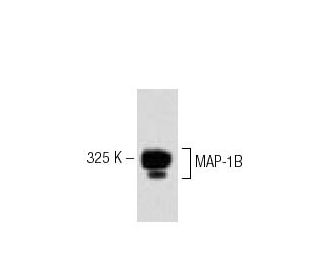
: sc-135978. Western blot analysis of MAP-1B expression in mouse fetus head tissue extract.")
MAP-1B Antibody (6): sc-135978
- MAP-1B Antibody (6) is a mouse monoclonal IgG2a, cited in 2 publications, provided at 50 µg/500 µl
- raised against amino acids 1745-1858 of MAP-1B of mouse origin
- recommended for detection of MAP-1B of mouse, rat and human origin by WB and IP
- At present, we have not yet completed the identification of the preferred secondary detection reagent(s) for MAP-1B Antibody (6). This work is in progress.

MAP-1B Antibody (6) is a mouse monoclonal IgG2a antibody that detects MAP-1B in mouse, rat, and human samples through western blotting (WB) and immunoprecipitation (IP) applications. MAP-1B is a crucial microtubule-associated protein that plays a significant role in the stabilization and assembly of microtubules, which are essential components of the cytoskeleton. MAP-1B (6) antibody is vital for maintaining cellular structure and facilitating intracellular transport, as MAP-1B helps regulate the dynamic instability of microtubules. This dynamic instability is critical for various cellular processes, including cell division and motility. Notably, MAP-1B interacts with tubulin, the building block of microtubules, and MAP-1B phosphorylation status can influence activity and interactions, thereby affecting microtubule dynamics. MAP-1B′s ability to modulate microtubule stability underscores its importance in cellular architecture and function, making MAP-1B a valuable target for research in neurobiology and cancer studies. Anti-MAP-1B antibody (6) is an essential tool for researchers aiming to explore the intricate roles of microtubule-associated proteins in health and disease.
Alexa Fluor® is a trademark of Molecular Probes Inc., OR., USA
LI-COR® and Odyssey® are registered trademarks of LI-COR Biosciences
MAP-1B Antibody (6) References:
- Acute inactivation of MAP1b in growing sympathetic neurons destabilizes axonal microtubules. | Tint, I., et al. 2005. Cell Motil Cytoskeleton. 60: 48-65. PMID: 15573412
- Cells positive for microtubule-associated protein 1B (MAP 1B) are present along rat and human efferent ductules and epididymis. | Queiróz, DB., et al. 2006. Cell Tissue Res. 325: 125-33. PMID: 16541288
- QKI binds MAP1B mRNA and enhances MAP1B expression during oligodendrocyte development. | Zhao, L., et al. 2006. Mol Biol Cell. 17: 4179-86. PMID: 16855020
- Quantitation of microtubule-associated protein MAP-1B in brain and other tissues. | Díaz-Nido, J. and Avila, J. 1989. Int J Biochem. 21: 723-30. PMID: 2759332
- MAP1B Light Chain Modulates Synaptic Transmission via AMPA Receptor Intracellular Trapping. | Palenzuela, R., et al. 2017. J Neurosci. 37: 9945-9963. PMID: 28904092
- MAP1B mutations cause intellectual disability and extensive white matter deficit. | Walters, GB., et al. 2018. Nat Commun. 9: 3456. PMID: 30150678
- Interaction of hnRNP K with MAP 1B-LC1 promotes TGF-β1-mediated epithelial to mesenchymal transition in lung cancer cells. | Li, L., et al. 2019. BMC Cancer. 19: 894. PMID: 31492158
- Mutations of MAP1B encoding a microtubule-associated phosphoprotein cause sensorineural hearing loss. | Cui, L., et al. 2020. JCI Insight. 5: PMID: 33268592
- Epilepsy phenotypes associated with MAP1B-related brain malformations. | Arya, R., et al. 2021. Epileptic Disord. 23: 392-396. PMID: 33772511
- Characterization of microtubule-associated protein MAP1B: phosphorylation state, light chains, and binding to microtubules. | Pedrotti, B., et al. 1996. Biochemistry. 35: 3016-23. PMID: 8608140
Ordering Information
| Product Name | Catalog # | UNIT | Price | Qty | FAVORITES | |
MAP-1B Antibody (6) | sc-135978 | 50 µg/500 µl | $322.00 |