
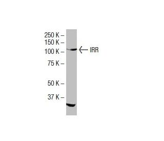


IRR Antibody (I-63): sc-100303
- IRR Antibody (I-63) is a mouse monoclonal IgG1 κ IRR antibody provided at 100 µg/ml
- raised against recombinant IRR of human origin
- recommended for detection of IRR of human origin by WB, IP, IF, IHC(P) and ELISA
- At present, we have not yet completed the identification of the preferred secondary detection reagent(s) for IRR Antibody (I-63). This work is in progress.

QUICK LINKS
IRR Antibody (I-63) is a mouse monoclonal IgG1 kappa light chain antibody that detects IRR protein of human origin by western blotting (WB), immunoprecipitation (IP), immunofluorescence (IF), immunohistochemistry with paraffin-embedded sections (IHCP), and enzyme-linked immunosorbent assay (ELISA). Anti-IRR antibody (I-63) is available as the non-conjugated isotype antibody. The insulin receptor-related receptor (IRR) plays a crucial role in cellular signaling pathways, particularly in insulin regulation, which is vital for maintaining glucose homeostasis in the body. Although the exact ligand for IRR remains unidentified, structural similarity to insulin and insulin-like growth factor-1 receptors suggests participation in similar signaling mechanisms. IRR is synthesized as a single polypeptide precursor that undergoes proteolytic cleavage and glycosylation, resulting in alpha and beta subunits that assemble into a heterotetramer. The two alpha subunits are responsible for ligand binding, while the beta subunits contain the kinase domain essential for downstream signaling. IRR is predominantly expressed in the kidney, with additional expression in the brain, stomach, pancreas, and heart, indicating potential involvement in various physiological processes. The gene encoding IRR is located on human chromosome 1q23.1, a region associated with type-2 diabetes mellitus, highlighting IRR′s importance in insulin signaling and metabolic regulation.
Alexa Fluor® is a trademark of Molecular Probes Inc., OR., USA
LI-COR® and Odyssey® are registered trademarks of LI-COR Biosciences
IRR Antibody (I-63) References:
- Preserved pancreatic beta-cell development and function in mice lacking the insulin receptor-related receptor. | Kitamura, T., et al. 2001. Mol Cell Biol. 21: 5624-30. PMID: 11463843
- Polymorphism screening of the insulin receptor-related receptor gene (INSRR) on 1q in Pima Indians. | Wolford, JK., et al. 2001. Mol Cell Probes. 15: 223-7. PMID: 11513557
- Insulin receptor-related receptor messenger ribonucleic acid: quantitative distribution and localization to subpopulations of epithelial cells in stomach and kidney. | Mathi, SK., et al. 1995. Endocrinology. 136: 4125-32. PMID: 7649121
- Expression of a cDNA encoding the human insulin receptor-related receptor. | Jui, HY., et al. 1994. J Biol Chem. 269: 22446-52. PMID: 8071374
- Localization of insulin receptor-related receptor in the rat kidney. | Ozaki, K., et al. 1997. Kidney Int. 52: 694-8. PMID: 9291189
- Insulin receptor-related receptor in rat islets of Langerhans. | Ozaki, K. 1998. Eur J Endocrinol. 139: 244-7. PMID: 9724084
- Effect of fasting on insulin receptor-related receptor messenger ribonucleic acid in rat kidney. | Chrysis, D., et al. 1998. J Endocrinol. 159: R9-R12. PMID: 9795378
Ordering Information
| Product Name | Catalog # | UNIT | Price | Qty | FAVORITES | |
IRR Antibody (I-63) | sc-100303 | 100 µg/ml | $339.00 |