
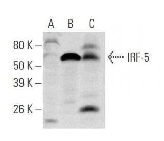


IRF-5 Anticuerpo (10T1): sc-56714
- IRF-5 Anticuerpo 10T1 es un monoclonal de ratón IgG1, ver las 14 publicaciones, proporcionado como 50 µg/0.5 ml
- producido contra los amino ácidos 176-240 de IRF-5 de origen human
- recomendado para detectarIRF-5 de mouse, rat y human origen, mediante WB, IP, IF y ELISA
- m-IgG Fc BP-HRP y m-IgG1 BP-HRP son los reactivos de detección secundarios preferidos para IRF-5 Anticuerpo (10T1) for WB applications. Estos reactivos se ofrecen ahora en paquetes con IRF-5 Anticuerpo (10T1)(véase la información de pedido más abajo).

El anticuerpo IRF-5 (10T1) es un anticuerpo monoclonal IgG1 de ratón que detecta el IRF-5 en muestras de ratón, rata y humanos a través de aplicaciones como el western blotting (WB), la inmunoprecipitación (IP), la inmunofluorescencia (IF) y el ensayo inmunoenzimático (ELISA). El anticuerpo IRF-5 (10T1) está disponible en forma no conjugada, lo que permite un uso experimental versátil. El factor regulador del interferón 5 (IRF-5) es un miembro crucial de la familia IRF de proteínas de unión al ADN, que también incluye el IRF-1, IRF-2, IRF-3, IRF-4, IRF-6, IRF-7, ISGF-3γ p48 y la proteína de unión a la secuencia consenso del interferón (ICSBP). Esta familia desempeña un papel importante en la regulación tanto de los genes inducibles por interferón como de los genes inducibles por interferón, lo que hace que el IRF-5 sea esencial para la modulación de la respuesta inmunitaria. En particular, el IRF-5 participa en las vías de señalización activadas por las infecciones víricas, transduce directamente señales que conducen a la expresión de diversas citocinas y quimiocinas, vitales para orquestar la respuesta inmunitaria. Además, se sabe que el IRF-5 interactúa con varias proteínas específicas, incluida la p53, que, aunque es una diana directa del IRF-5, no influye en las funciones reguladoras del ciclo celular y pro-apoptóticas, lo que pone de relieve los complejos mecanismos reguladores en juego en la señalización inmunitaria y las respuestas celulares.
Alexa Fluor® es una marca registrada de Molecular Probes Inc., OR., USA
REIVEW LI-COR® y Odyssey® son marcas registradas de LI-COR Biosciences.
IRF-5 Anticuerpo (10T1) Referencias:
- Factores reguladores del interferón: la próxima generación. | Mamane, Y., et al. 1999. Gene. 237: 1-14. PMID: 10524230
- La activación específica del virus de un nuevo factor regulador del interferón, IRF-5, da lugar a la inducción de distintos genes de interferón alfa. | Barnes, BJ., et al. 2001. J Biol Chem. 276: 23382-90. PMID: 11303025
- Identificación del gen del factor regulador del interferón 5 (IRF-5) como diana directa de p53. | Mori, T., et al. 2002. Oncogene. 21: 2914-8. PMID: 11973653
- Múltiples dominios reguladores de IRF-5 controlan la activación, la localización celular y la inducción de quimiocinas que median en el reclutamiento de linfocitos T. | Barnes, BJ., et al. 2002. Mol Cell Biol. 22: 5721-40. PMID: 12138184
- La formación de heterodímeros inducida por virus entre IRF-5 e IRF-7 modula el ensamblaje del enhanceosome IFNA in vivo y la actividad transcripcional de los genes IFNA. | Barnes, BJ., et al. 2003. J Biol Chem. 278: 16630-41. PMID: 12600985
- El factor regulador del interferón 5, un nuevo mediador de la detención del ciclo celular y la muerte celular. | Barnes, BJ., et al. 2003. Cancer Res. 63: 6424-31. PMID: 14559832
- Contribución única del eje IRF-5-Ikaros a la respuesta IgG2a de las células B. | Fang, CM., et al. 2012. Genes Immun. 13: 421-30. PMID: 22535200
- IRF-5 y NF-κB p50 co-regulan la expresión de IFN-β e IL-6 en células dendríticas plasmocitoides humanas estimuladas por TLR9. | Steinhagen, F., et al. 2013. Eur J Immunol. 43: 1896-906. PMID: 23616277
- Expresión constitutiva de IRF-5 en células T infectadas por HTLV-1. | Ishikawa, C., et al. 2015. Int J Oncol. 47: 361-9. PMID: 26004104
- IRF-5 promueve la muerte celular en células T CD4 durante la infección crónica. | Fabié, A., et al. 2018. Cell Rep. 24: 1163-1175. PMID: 30067973
- El eje TLR7/IRF-5 sensibiliza las células T CD4+ de memoria a la apoptosis mediada por Fas durante la infección por VIH-1. | Carmona-Pérez, L., et al. 2023. JCI Insight. 8: PMID: 37227774
- Vías Jak-STAT y activación transcripcional en respuesta a IFNs y otras proteínas de señalización extracelular. | Darnell, JE., et al. 1994. Science. 264: 1415-21. PMID: 8197455
Información sobre pedidos
| Nombre del producto | Número de catálogo | UNIDAD | Precio | CANTIDAD | Favoritos | |
IRF-5 Anticuerpo (10T1) | sc-56714 | 50 µg/0.5 ml | $322.00 | |||
Paquete de IRF-5 (10T1): m-IgG Fc BP-HRP | sc-539038 | 50 µg Ab; 10 µg BP | $361.00 | |||
Paquete de IRF-5 (10T1): m-IgG1 BP-HRP | sc-541221 | 50 µg Ab; 20 µg BP | $361.00 |