")
ILKAP Antibody (41): sc-136341
- ILKAP Antibody (41) is a mouse monoclonal IgG1 κ ILKAP antibody provided at 200 µg/ml
- raised against amino acids 38-144 of ILKAP of rat origin
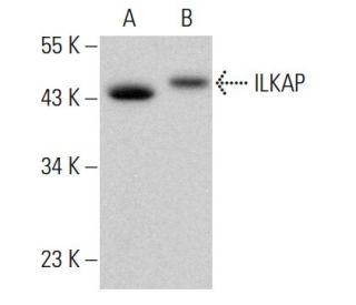
- ILKAP Antibody (41) is recommended for detection of ILKAP of mouse, rat and human origin by WB, IP and IF
- ILKAP Antibody (41) is available conjugated to agarose for IP; and to HRP for WB, IHC(P) and ELISA
- m-IgG Fc BP-HRP, m-IgG1 BP-HRP and m-IgGκ BP-HRP are the preferred secondary detection reagents for ILKAP Antibody (41) for WB applications. These reagents are now offered in bundles with ILKAP Antibody (41) (see ordering information below).

QUICK LINKS
SEE ALSO...
ILKAP Antibody (41) is a mouse monoclonal IgG1 kappa light chain antibody that detects ILKAP protein of mouse, rat, and human origin by western blotting (WB), immunoprecipitation (IP), and immunofluorescence (IF). Anti-ILKAP antibody (41) is available in both non-conjugated and various conjugated forms, including agarose and horseradish peroxidase (HRP). ILKAP, also known as integrin-linked kinase-associated serine/threonine phosphatase 2C or PP2C-delta, is a 392 amino acid cytoplasmic protein phosphatase that plays a crucial role in regulating growth factor signaling and cell adhesion by selectively interacting with integrin-linked kinase (ILK). This interaction is vital for maintaining cellular functions and ensuring proper responses to extracellular signals, which can influence processes such as cell migration, proliferation, and survival. ILKAP is predominantly expressed in striated muscle, with lower expression levels in smooth muscle, highlighting its importance in muscle physiology. As a member of the PP2C family, ILKAP contains a PP2C-like domain and binds two magnesium or manganese ions per subunit as cofactors, which are essential for phosphatase activity. The gene encoding ILKAP is located on human chromosome 2, a region that encompasses 237 million bases and encodes over 1,400 genes, representing approximately 8% of the human genome.
Alexa Fluor® is a trademark of Molecular Probes Inc., OR., USA
LI-COR® and Odyssey® are registered trademarks of LI-COR Biosciences
ILKAP Antibody (41) References:
- Modulation of integrin signal transduction by ILKAP, a protein phosphatase 2C associating with the integrin-linked kinase, ILK1. | Leung-Hagesteijn, C., et al. 2001. EMBO J. 20: 2160-70. PMID: 11331582
- ILKAP regulates ILK signaling and inhibits anchorage-independent growth. | Kumar, AS., et al. 2004. Oncogene. 23: 3454-61. PMID: 14990992
- PP2C family members play key roles in regulation of cell survival and apoptosis. | Tamura, S., et al. 2006. Cancer Sci. 97: 563-7. PMID: 16827794
- Expression of stress inducible protein 1 (Stip1) in the mouse testis. | Mizrak, SC., et al. 2006. Mol Reprod Dev. 73: 1361-6. PMID: 16894550
- Role of type 2C protein phosphatases in growth regulation and in cellular stress signaling. | Lammers, T. and Lavi, S. 2007. Crit Rev Biochem Mol Biol. 42: 437-61. PMID: 18066953
- Modulation of integrin-linked kinase nucleo-cytoplasmic shuttling by ILKAP and CRM1. | Nakrieko, KA., et al. 2008. Cell Cycle. 7: 2157-66. PMID: 18635968
- Proteomic analysis reveals Hrs ubiquitin-interacting motif-mediated ubiquitin signaling in multiple cellular processes. | Pridgeon, JW., et al. 2009. FEBS J. 276: 118-31. PMID: 19019082
- Stress-induced phosphoprotein 1 restrains spinal cord ischaemia-reperfusion injury by modulating NF-κB signalling. | Jin, H., et al. 2021. J Cell Mol Med. 25: 11075-11084. PMID: 34734476
- Intracellular targeting of STIP1 inhibits human cancer cell line growth. | Lin, CY., et al. 2021. Transl Cancer Res. 10: 1313-1323. PMID: 35116457
- Cross-talk between BCKDK-mediated phosphorylation and STUB1-dependent ubiquitination degradation of BCAT1 promotes GBM progression. | Wang, W., et al. 2024. Cancer Lett. 591: 216849. PMID: 38621458
- Proteostasis perturbation of N-Myc leveraging HSP70 mediated protein turnover improves treatment of neuroendocrine prostate cancer. | Xu, P., et al. 2024. Nat Commun. 15: 6626. PMID: 39103353
- Cloning and characterization of a novel mammalian PP2C isozyme. | Tong, Y., et al. 1998. J Biol Chem. 273: 35282-90. PMID: 9857069
Ordering Information
| Product Name | Catalog # | UNIT | Price | Qty | FAVORITES | |
ILKAP Antibody (41) | sc-136341 | 200 µg/ml | $322.00 | |||
ILKAP Antibody (41): m-IgG Fc BP-HRP Bundle | sc-528814 | 200 µg Ab; 10 µg BP | $361.00 | |||
ILKAP Antibody (41): m-IgGκ BP-HRP Bundle | sc-521358 | 200 µg Ab, 40 µg BP | $361.00 | |||
ILKAP Antibody (41): m-IgG1 BP-HRP Bundle | sc-543145 | 200 µg Ab; 20 µg BP | $361.00 | |||
ILKAP Antibody (41) AC | sc-136341 AC | 500 µg/ml, 25% agarose | $424.00 | |||
ILKAP Antibody (41) HRP | sc-136341 HRP | 200 µg/ml | $322.00 |