")
Anticorps Glycophorin A (R10): sc-53905
- L'anticorps Glycophorin A R10 est un monoclonal IgG1 κ L'Anticorps Glycophorin A, cité dans 3 publications, fourni en 200 µg/ml
- élevé contre des cellules tumorales pré-B induites par le virus de la leucémie murine d'Abelson human.
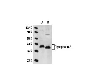
- L'Anticorps Glycophorin A (R10) est recommandé pour la détection de Glycophorin A d'origine human par WB, IP, IF, IHC(P) et FCM
- Anti-L'Anticorps Glycophorin A (R10) est disponible conjugué à l'agarose pour IP; à l'HRP pour WB, IHC(P) et ELISA; et soit à la phycoerythrin ou FITC pour IF, IHC(P) et FCM
- aussi disponible conjugué à l'Alexa Fluor® 488, Alexa Fluor® 546, Alexa Fluor® 594 ou Alexa Fluor® 647 pour WB (RGB), IF, IHC(P) et FCM
- aussi disponible conjugué à l'Alexa Fluor® 680 ou Alexa Fluor® 790 pour WB (NIR), IF et FCM
- Contactez notre Service Technique (ou votre Distributeur local) pour plus d'informations pour recevoir un échantillon GRATUIT de 10 µg de Glycophorin A (R10): sc-53905.
- m-IgG Fc BP-HRP, m-IgG1 BP-HRP et m-IgGκ BP-HRP sont les réactifs secondaires recommandés pour la détection de l'anticorps Glycophorin A (R10) pour des applications de WB and IHC(P). Ces réactifs sont maintenant proposés en kit avec l'anticorps Glycophorin A (R10) (voir Informations pour la commande ci-dessous).
ACCÈS RAPIDE AUX LIENS
VOIR ÉGALEMENT...
L'anticorps anti-glycophorine A (R10) est un anticorps monoclonal de souris IgG1 à chaîne légère kappa qui détecte la glycophorine A d'origine humaine par western blotting (WB), immunoprécipitation (IP), immunofluorescence (IF), immunohistochimie sur coupes incluses en paraffine (IHCP) et cytométrie de flux (FCM). L'anticorps anti-glycophorine A (R10) est disponible sous forme non conjuguée et sous diverses formes conjuguées, notamment agarose, peroxydase de raifort (HRP), phycoérythrine (PE), isothiocyanate de fluorescéine (FITC) et plusieurs conjugués Alexa Fluor®. La glycophorine A est une sialoglycoprotéine cruciale située à la surface des érythrocytes humains, qui joue un rôle important dans le maintien de l'intégrité structurelle des globules rouges et facilite l'interaction avec le système immunitaire. La glycophorine A traverse la membrane une fois, présentant une extrémité amino-terminale à l'environnement extracellulaire, ce qui est essentiel pour la fonction d'antigénicité du groupe sanguin. La diversité génétique des antigènes de surface des glycophorines, y compris les glycophorines A, B et C, détermine le phénotype du groupe sanguin, ce qui rend ces protéines vitales pour la compatibilité des transfusions sanguines et la compréhension des maladies hémolytiques. Le gène de la Glycophorine A, situé sur le chromosome 4q31.21, se compose de sept exons et présente une forte homologie avec la Glycophorine B, codant pour une protéine de 91 acides aminés. La compréhension de la structure et de la fonction de la glycophorine A reste cruciale pour les applications cliniques et la recherche en biologie érythrocytaire, car elle permet de mieux comprendre l'expression des antigènes de groupe sanguin et les cibles thérapeutiques potentielles pour les troubles liés au sang.
Alexa Fluor® est une marque déposée de Molecular Probes Inc., OR., USA
LI-COR® et Odyssey® sont marques déposées de LI-COR Biosciences
Anticorps Glycophorin A (R10) Références:
- Identification immunohistochimique des précurseurs érythroïdes dans des coupes de moelle osseuse incluses en paraffine: la spectrine est un marqueur supérieur à la glycophorine. | Sadahira, Y., et al. 1999. J Clin Pathol. 52: 919-21. PMID: 10711257
- Détection in vivo de l'hétéro-association de la glycophorine-A et de ses mutants dans la membrane. | Gerber, D. and Shai, Y. 2001. J Biol Chem. 276: 31229-32. PMID: 11402026
- Des régions distinctes de la glycophorine A humaine améliorent la fonction de transport et le trafic de surface de l'échangeur d'anions des globules rouges humains (bande 3; AE1). | Young, MT. and Tanner, MJ. 2003. J Biol Chem. 278: 32954-61. PMID: 12813056
- Exigence de la glycophorine A pour l'expression d'antigènes liés à l'oxygène sur la membrane des érythrocytes. | Arimitsu, N., et al. 2003. Genes Cells. 8: 769-77. PMID: 12940824
- Structure altérée et propriétés de transport d'anions de la bande 3 (AE1, SLC4A1) dans les globules rouges humains dépourvus de glycophorine A. | Bruce, LJ., et al. 2004. J Biol Chem. 279: 2414-20. PMID: 14604989
- Adaptation structurelle de l'homodimère transmembranaire de la glycophorine A aux modifications de l'acide aminé D. | Gerber, D., et al. 2004. J Mol Biol. 339: 243-50. PMID: 15123435
- Des interactions complexes à l'interface hélice-hélice stabilisent le dimère transmembranaire de la glycophorine A. | Doura, AK. and Fleming, KG. 2004. J Mol Biol. 343: 1487-97. PMID: 15491626
- La co-transfection de NXPE2 murine et de glycophorine A murine confère une réactivité avec Ter-119. | Keele, GR., et al. 2024. Haematologica. 109: 3755-3759. PMID: 39021224
- La glycophorine A comme marqueur de surface cellulaire de la différenciation érythroïde précoce dans la leucémie aiguë. | Andersson, LC., et al. 1979. Int J Cancer. 24: 717-20. PMID: 397196
- Expression de la glycophorine A dans l'hématopoïèse maligne. | Liszka, K., et al. 1983. Am J Hematol. 15: 219-26. PMID: 6638008
- Expression des antigènes de surface cellulaire dans les progéniteurs érythroïdes humains: marqueurs érythroïdes et mégacaryocytaires. | Nakahata, T. and Okumura, N. 1994. Leuk Lymphoma. 13: 401-9. PMID: 8069185
- Différenciation entre les ecchymoses et les décolorations putréfactives de la peau par analyse immunologique de la glycophorine A. | Kibayashi, K., et al. 1993. Forensic Sci Int. 61: 111-7. PMID: 8307520
Informations pour la commande
| Nom du produit | Ref. Catalogue | COND. | Prix HT | QTÉ | Favoris | |
Anticorps Glycophorin A (R10) | sc-53905 | 200 µg/ml | $316.00 | |||
Glycophorin A (R10): m-IgG Fc BP-HRP Kit | sc-528587 | 200 µg Ab; 10 µg BP | $354.00 | |||
Glycophorin A (R10): m-IgGκ BP-HRP Kit | sc-520999 | 200 µg Ab, 40 µg BP | $354.00 | |||
Glycophorin A (R10): m-IgG1 BP-HRP Kit | sc-543011 | 200 µg Ab; 20 µg BP | $354.00 | |||
Anticorps Glycophorin A (R10) AC | sc-53905 AC | 500 µg/ml, 25% agarose | $416.00 | |||
Anticorps Glycophorin A (R10) HRP | sc-53905 HRP | 200 µg/ml | $316.00 | |||
Anticorps Glycophorin A (R10) FITC | sc-53905 FITC | 200 µg/ml | $330.00 | |||
Anticorps Glycophorin A (R10) PE | sc-53905 PE | 200 µg/ml | $343.00 | |||
Anticorps Glycophorin A (R10) Alexa Fluor® 488 | sc-53905 AF488 | 200 µg/ml | $357.00 | |||
Anticorps Glycophorin A (R10) Alexa Fluor® 546 | sc-53905 AF546 | 200 µg/ml | $357.00 | |||
Anticorps Glycophorin A (R10) Alexa Fluor® 594 | sc-53905 AF594 | 200 µg/ml | $357.00 | |||
Anticorps Glycophorin A (R10) Alexa Fluor® 647 | sc-53905 AF647 | 200 µg/ml | $357.00 | |||
Anticorps Glycophorin A (R10) Alexa Fluor® 680 | sc-53905 AF680 | 200 µg/ml | $357.00 | |||
Anticorps Glycophorin A (R10) Alexa Fluor® 790 | sc-53905 AF790 | 200 µg/ml | $357.00 |