")
: sc-66192. Immunoperoxidase staining of formalin fixed, paraffin-embedded human vulva/anal skin tissue showing staining of stratum corneum.")
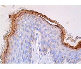
: sc-66192. Immunoperoxidase staining of formalin fixed, paraffin-embedded human skin tissue showing staining of stratum corneum.")
FLG/Filaggrin Antibody (AKH1): sc-66192
- FLG/Filaggrin Antibody (AKH1) is a mouse monoclonal IgG1 κ FLG/Filaggrin antibody, cited in 98 publications, provided at 200 µg/ml
- raised against purified foreskin Filaggrin of human origin
- FLG/Filaggrin Antibody (AKH1) is recommended for detection of Filaggrin and Profilaggrin of human origin by WB, IF and IHC(P)
- Anti-FLG/Filaggrin Antibody (AKH1) is available conjugated to agarose for IP; HRP for WB, IHC(P) and ELISA; and to either phycoerythrin or FITC for IF, IHC(P) and FCM
- also available conjugated to Alexa Fluor® 488, Alexa Fluor® 546, Alexa Fluor® 594 or Alexa Fluor® 647 for WB (RGB), IF, IHC(P) and FCM, and for use with RGB fluorescent imaging systems, such as iBright™ FL1000, FluorChem™, Typhoon, Azure and other comparable systems
- also available conjugated to Alexa Fluor® 680 or Alexa Fluor® 790 for WB (NIR), IF and FCM; for use with Near-Infrared (NIR) detection systems, such as LI-COR®Odyssey®, iBright™ FL1000, FluorChem™, Typhoon, Azure and other comparable systems
- m-IgG Fc BP-HRP and m-IgG1 BP-HRP are the preferred secondary detection reagents for FLG/Filaggrin Antibody (AKH1) for WB and IHC(P) applications. These reagents are now offered in bundles with FLG/Filaggrin Antibody (AKH1) (see ordering information below).

QUICK LINKS
SEE ALSO...
Filaggrin Antibody (AKH1) is a mouse monoclonal IgG1 kappa light chain antibody that detects Filaggrin protein of human origin by western blotting (WB), immunofluorescence (IF), and immunohistochemistry with paraffin-embedded sections (IHCP). Anti-Filaggrin antibody (AKH1) is available in both non-conjugated and various conjugated forms, including agarose, horseradish peroxidase (HRP), phycoerythrin (PE), fluorescein isothiocyanate (FITC), and multiple Alexa Fluor® conjugates, providing versatility for different experimental needs. Filaggrin, a key structural protein, plays a crucial role in the skin′s barrier function by promoting the aggregation of keratin intermediate filaments, which is essential for maintaining the integrity and resilience of the epidermis. Filaggrin is primarily located in the keratohyalin granules of the epidermis, where synthesis occurs as a large precursor known as profilaggrin. During the terminal differentiation of keratinocytes, profilaggrin undergoes proteolytic cleavage into active filaggrin, which facilitates the formation of a robust cornified envelope that protects against environmental stressors and prevents water loss. Proper functioning of filaggrin is vital for skin health, as mutations in the filaggrin gene are associated with various skin disorders, including atopic dermatitis and ichthyosis vulgaris, highlighting filaggrin′s importance in both skin barrier function and overall dermatological health.
Alexa Fluor® is a trademark of Molecular Probes Inc., OR., USA
LI-COR® and Odyssey® are registered trademarks of LI-COR Biosciences
FLG/Filaggrin Antibody (AKH1) References:
- Inducible expression of filaggrin increases keratinocyte susceptibility to apoptotic cell death. | Kuechle, MK., et al. 2000. Cell Death Differ. 7: 566-73. PMID: 10822280
- Filaggrin mutations associated with skin and allergic diseases. | Irvine, AD., et al. 2011. N Engl J Med. 365: 1315-27. PMID: 21991953
- Organization, structure, and polymorphisms of the human profilaggrin gene. | Gan, SQ., et al. 1990. Biochemistry. 29: 9432-40. PMID: 2248957
- Filaggrin and Skin Barrier Function. | Kezic, S. and Jakasa, I. 2016. Curr Probl Dermatol. 49: 1-7. PMID: 26844893
- Characterization of a cDNA clone encoding human filaggrin and localization of the gene to chromosome region 1q21. | McKinley-Grant, LJ., et al. 1989. Proc Natl Acad Sci U S A. 86: 4848-52. PMID: 2740331
- The role of filaggrin in atopic dermatitis and allergic disease. | Drislane, C. and Irvine, AD. 2020. Ann Allergy Asthma Immunol. 124: 36-43. PMID: 31622670
- Revisiting the Roles of Filaggrin in Atopic Dermatitis. | Moosbrugger-Martinz, V., et al. 2022. Int J Mol Sci. 23: PMID: 35628125
- PDE4 inhibition by difamilast regulates filaggrin and loricrin expression via keratinocyte proline-rich protein in human keratinocytes. | Tsuji, G., et al. 2023. J Dermatol Sci. 110: 61-68. PMID: 37156706
- Influence of pathogenic filaggrin variants on dupilumab treatment in atopic dermatitis. | Clabbers, J., et al. 2024. J Allergy Clin Immunol. 153: 1155-1161.e4. PMID: 38272373
- Filaggrin Mutation Status and Prevention of Atopic Dermatitis with Maternal Probiotic Supplementation. | Zakiudin, DP., et al. 2024. Acta Derm Venereol. 104: adv24360. PMID: 38655655
- Filaggrin linker segment peptide and cystatin alpha are parts of a complex of the cornified envelope of epidermis. | Takahashi, M., et al. 1996. Arch Biochem Biophys. 329: 123-6. PMID: 8619628
- Recruitment of cycling epidermal cells and expression of filaggrin, involucrin and tenascin in the margin of the active psoriatic plaque, in the uninvolved skin of psoriatic patients and in the normal healthy skin. | Gerritsen, MJ., et al. 1997. J Dermatol Sci. 14: 179-88. PMID: 9138475
Ordering Information
| Product Name | Catalog # | UNIT | Price | Qty | FAVORITES | |
FLG/Filaggrin Antibody (AKH1) | sc-66192 | 200 µg/ml | $322.00 | |||
FLG/Filaggrin Antibody (AKH1): m-IgG Fc BP-HRP Bundle | sc-526977 | 200 µg Ab; 10 µg BP | $361.00 | |||
FLG/Filaggrin Antibody (AKH1): m-IgG1 BP-HRP Bundle | sc-532350 | 200 µg Ab; 20 µg BP | $361.00 | |||
FLG/Filaggrin Antibody (AKH1) AC | sc-66192 AC | 500 µg/ml, 25% agarose | $424.00 | |||
FLG/Filaggrin Antibody (AKH1) HRP | sc-66192 HRP | 200 µg/ml | $322.00 | |||
FLG/Filaggrin Antibody (AKH1) FITC | sc-66192 FITC | 200 µg/ml | $336.00 | |||
FLG/Filaggrin Antibody (AKH1) PE | sc-66192 PE | 200 µg/ml | $349.00 | |||
FLG/Filaggrin Antibody (AKH1) Alexa Fluor® 488 | sc-66192 AF488 | 200 µg/ml | $364.00 | |||
FLG/Filaggrin Antibody (AKH1) Alexa Fluor® 546 | sc-66192 AF546 | 200 µg/ml | $364.00 | |||
FLG/Filaggrin Antibody (AKH1) Alexa Fluor® 594 | sc-66192 AF594 | 200 µg/ml | $364.00 | |||
FLG/Filaggrin Antibody (AKH1) Alexa Fluor® 647 | sc-66192 AF647 | 200 µg/ml | $364.00 | |||
FLG/Filaggrin Antibody (AKH1) Alexa Fluor® 680 | sc-66192 AF680 | 200 µg/ml | $364.00 | |||
FLG/Filaggrin Antibody (AKH1) Alexa Fluor® 790 | sc-66192 AF790 | 200 µg/ml | $364.00 |