")
E-cadherin Antikörper (G-10): sc-8426
- E-cadherin Antikörper G-10 ist ein Maus monoklonales IgG1 κ E-cadherin Antikörper, verwendet in 629 wissenschaftlichen Veröffentlichungen, in einer Menge von 200 µg/ml
- gegen Aminosäuren 600-707 gerichtet, Kartierung innerhalb der extrazellulären Domäne von E-cadherin mit Ursprung human
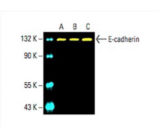
- E-cadherin Antikörper (G-10) ist empfohlen für die Detektion von E-cadherin aus der Spezies mouse, rat und human per WB, IP, IF, IHC(P) und ELISA
- Anti-E-cadherin Antikörper (G-10) ist erhältlich als Konjugat mit Agarose für IP; HRP für WB, IHC(P) und ELISA; und entweder mit Phycoerythrin oder FITC für IF, IHC(P) und FCM
- auch erhältlich als Konjugat mit Alexa Fluor® 488, Alexa Fluor® 546, Alexa Fluor® 594 oder Alexa Fluor® 647 für IF, IHC(P) und FCM
- auch erhältlich als Konjugat mit Alexa Fluor® 680 oder Alexa Fluor® 790 für WB (NIR), IF und FCM
- m-IgG Fc BP-HRP ist das bevorzugte sekundäre Nachweisreagenz für E-cadherin Antikörper (G-10) für WB- und IHC(P)-Anwendungen. Dieses Reagenz wird jetzt in einem Paket mit E-cadherin Antikörper (G-10) angeboten(siehe Bestellinformationen unten).

Direktverknüpfungen
Siehe auch...
Der E-Cadherin-Antikörper (G-10) ist ein monoklonaler IgG1-Antikörper der leichten Kette von Mäusen, der E-Cadherin-Protein von Mäusen, Ratten und Menschen durch Western Blot (WB), Immunopräzipitation (IP), Immunfluoreszenz (IF), Immunhistochemie und Enzyme-linked Immunosorbent Assay (ELISA) nachweist. Der Anti-E-Cadherin-Antikörper (G-10) ist sowohl in nicht konjugierter als auch in verschiedenen konjugierten Formen erhältlich, darunter Agarose, Meerrettichperoxidase (HRP), Phycoerythrin (PE), Fluorescein-Isothiocyanat (FITC) und mehrere Alexa Fluor®-Konjugate. E-Cadherin, auch bekannt als CD324 oder CDH1, spielt eine entscheidende Rolle bei der Zelladhäsion, die für die Aufrechterhaltung der Gewebeintegrität und die Erleichterung morphogenetischer Prozesse während der Embryonalentwicklung von entscheidender Bedeutung ist. Das E-Cadherin-Protein befindet sich hauptsächlich an der Zellmembran, wo E-Cadherin mit benachbarten Zellen Adhäsionsstellen bildet und so zur strukturellen Stabilität des Gewebes beiträgt. Die Anwesenheit von E-Cadherin an diesen Stellen ist für die Regulierung der zellulären Signalwege und die Aufrechterhaltung der epithelialen Zellpolarität unerlässlich. Eine Störung der E-Cadherin-Funktion wird mit verschiedenen pathologischen Zuständen in Verbindung gebracht, darunter Krebsmetastasen, bei denen der Verlust der Zelladhäsion eine erhöhte Zellmotilität und -invasion ermöglicht. Die Interaktion von E-Cadherin mit zytoplasmatischen Proteinen wie β-Catenin unterstreicht die Bedeutung von E-Cadherin für die zelluläre Kommunikation und Signalübertragung und macht den monoklonalen E-Cadherin-Antikörper (G-10) zu einem wertvollen Instrument für die Untersuchung dieser kritischen biologischen Prozesse.
Alexa Fluor® ist ein Markenzeichen von Molecular Probes Inc., OR., USA
LI-COR® und Odyssey® sind Markenzeichen von LI-COR Biosciences
E-cadherin Antikörper (G-10) Literaturhinweise:
- Die Expression des zellulären Adhäsionsmoleküls E-Cadherin ist bei hochgradigem Prostatakrebs reduziert oder nicht vorhanden. | Umbas, R., et al. 1992. Cancer Res. 52: 5104-9. PMID: 1516067
- Genomische Organisation und chromosomale Kartierung des P-Cadherin-Gens der Maus. | Hatta, M., et al. 1991. Nucleic Acids Res. 19: 4437-41. PMID: 1886768
- [Chirurgische Therapie von Brustkrebs]. | Hirsch, HA. 1978. Gynakol Rundsch. 18: 132-41. PMID: 207622
- Die Cadherine: Zell-Zell-Adhäsionsmoleküle, die die Morphogenese von Tieren steuern. | Takeichi, M. 1988. Development. 102: 639-55. PMID: 3048970
- Desmosomale Cadherine: eine weitere wachsende Multigenfamilie von Adhäsionsmolekülen. | Koch, PJ. and Franke, WW. 1994. Curr Opin Cell Biol. 6: 682-7. PMID: 7833048
- Cadherine und Catenine: Interaktionen und Funktionen in der Embryonalentwicklung. | Ranscht, B. 1994. Curr Opin Cell Biol. 6: 740-6. PMID: 7833053
- Die Klonierung von fünf menschlichen Cadherinen klärt charakteristische Merkmale der extrazellulären Domäne von Cadherinen und liefert weitere Beweise für zwei strukturell unterschiedliche Cadherin-Typen. | Tanihara, H., et al. 1994. Cell Adhes Commun. 2: 15-26. PMID: 7982033
- Räumliche und zeitliche Beziehungen zwischen Cadherinen und PECAM-1 in Zell-Zell-Verbindungen von menschlichen Endothelzellen. | Ayalon, O., et al. 1994. J Cell Biol. 126: 247-58. PMID: 8027182
- Dynamik der Bildung des Cadherin/Catenin-Komplexes: neue Proteininteraktionen und Wege der Komplexbildung. | Hinck, L., et al. 1994. J Cell Biol. 125: 1327-40. PMID: 8207061
Bestellinformation
| Produkt | Katalog # | EINHEIT | Preis | ANZAHL | Favoriten | |
E-cadherin Antikörper (G-10) | sc-8426 | 200 µg/ml | $322.00 | |||
E-cadherin (G-10): m-IgG Fc BP-HRP Bundle | sc-525454 | 200 µg Ab; 10 µg BP | $361.00 | |||
E-cadherin Antikörper (G-10) AC | sc-8426 AC | 500 µg/ml, 25% agarose | $424.00 | |||
E-cadherin Antikörper (G-10) HRP | sc-8426 HRP | 200 µg/ml | $322.00 | |||
E-cadherin Antikörper (G-10) FITC | sc-8426 FITC | 200 µg/ml | $336.00 | |||
E-cadherin Antikörper (G-10) PE | sc-8426 PE | 200 µg/ml | $349.00 | |||
E-cadherin Antikörper (G-10) Alexa Fluor® 488 | sc-8426 AF488 | 200 µg/ml | $364.00 | |||
E-cadherin Antikörper (G-10) Alexa Fluor® 546 | sc-8426 AF546 | 200 µg/ml | $364.00 | |||
E-cadherin Antikörper (G-10) Alexa Fluor® 594 | sc-8426 AF594 | 200 µg/ml | $364.00 | |||
E-cadherin Antikörper (G-10) Alexa Fluor® 647 | sc-8426 AF647 | 200 µg/ml | $364.00 | |||
E-cadherin Antikörper (G-10) Alexa Fluor® 680 | sc-8426 AF680 | 200 µg/ml | $364.00 | |||
E-cadherin Antikörper (G-10) Alexa Fluor® 790 | sc-8426 AF790 | 200 µg/ml | $364.00 |