")
DBC-1 Antibody (H-2): sc-166733
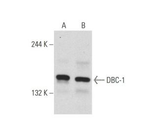
- DBC-1 Antibody (H-2) is a mouse monoclonal IgG3 κ DBC-1 antibody, cited in 5 publications, provided at 200 µg/ml
- specific for an epitope mapping between amino acids 750-768 within an internal region of DBC-1 of human origin
- recommended for detection of DBC-1 of human origin, 2610301G19Rik of mouse origin and the corresponding rat homolog by WB, IP, IF, IHC(P) and ELISA
- m-IgG3 BP-HRP and m-IgGκ BP-HRP are the preferred secondary detection reagents for DBC-1 Antibody (H-2) for WB and IHC(P) applications. These reagents are now offered in bundles with DBC-1 Antibody (H-2) (see ordering information below).

DBC-1 Antibody (H-2) is a mouse monoclonal IgG3 kappa light chain antibody that detects the DBC-1 protein of human origin, the 2610301G19Rik protein of mouse origin, and the corresponding rat homolog by western blotting (WB), immunoprecipitation (IP), immunofluorescence (IF), immunohistochemistry with paraffin-embedded sections (IHCP), and enzyme-linked immunosorbent assay (ELISA). Anti-DBC-1 antibody (H-2) is available as a non-conjugated format. DBC-1, also known as deleted in breast cancer gene 1 protein or p30 DBC protein, plays a critical role in cellular processes, particularly in the context of cancer biology. Located on chromosome 8 (8p21-8p23), DBC-1 is often found to be homozygously deleted in certain breast cancers, highlighting its importance as a tumor suppressor. DBC-1 features a nuclear localization signal, an N-terminal leucine zipper, an EF hand, and a C-terminal coiled-coil region, which are essential for its function in regulating apoptosis and cell growth. Notably, DBC-1 undergoes caspase-dependent processing during death signaling mediated by TNFα, resulting in the generation of DBC-1 p120 and p66 forms that translocate to the cytoplasm. DBC-1 p120 overexpression has been shown to induce mitochondrial clustering and enhance the sensitivity of cells to TNFα-mediated apoptosis. Furthermore, DBC-1 directly interacts with unliganded estrogen receptor alpha (ERα), stabilizing ERα expression and thereby contributing to the suppression of apoptosis and the promotion of hormone-independent cell proliferation. This multifaceted role of DBC-1 underscores its significance in cancer progression and therapeutic resistance, making DBC-1 (H-2) antibody a valuable tool for research in cancer biology and related fields.
Alexa Fluor® is a trademark of Molecular Probes Inc., OR., USA
LI-COR® and Odyssey® are registered trademarks of LI-COR Biosciences
DBC-1 Antibody (H-2) References:
- DBC2, a candidate for a tumor suppressor gene involved in breast cancer. | Hamaguchi, M., et al. 2002. Proc Natl Acad Sci U S A. 99: 13647-52. PMID: 12370419
- Comprehensive whole genome array CGH profiling of mantle cell lymphoma model genomes. | de Leeuw, RJ., et al. 2004. Hum Mol Genet. 13: 1827-37. PMID: 15229187
- Caspase-dependent processing activates the proapoptotic activity of deleted in breast cancer-1 during tumor necrosis factor-alpha-mediated death signaling. | Sundararajan, R., et al. 2005. Oncogene. 24: 4908-20. PMID: 15824730
- Characterization of 8p21.3 chromosomal deletions in B-cell lymphoma: TRAIL-R1 and TRAIL-R2 as candidate dosage-dependent tumor suppressor genes. | Rubio-Moscardo, F., et al. 2005. Blood. 106: 3214-22. PMID: 16051735
- Pressure stimulates breast cancer cell adhesion independently of cell cycle and apoptosis regulatory protein (CARP)-1 regulation of focal adhesion kinase. | Downey, C., et al. 2006. Am J Surg. 192: 631-5. PMID: 17071197
- Large-scale identification of c-MYC-associated proteins using a combined TAP/MudPIT approach. | Koch, HB., et al. 2007. Cell Cycle. 6: 205-17. PMID: 17314511
- Modulation of estrogen receptor alpha protein level and survival function by DBC-1. | Trauernicht, AM., et al. 2007. Mol Endocrinol. 21: 1526-36. PMID: 17473282
- Genomic assessments of the frequent loss of heterozygosity region on 8p21.3-p22 in head and neck squamous cell carcinoma. | Ye, H., et al. 2007. Cancer Genet Cytogenet. 176: 100-6. PMID: 17656251
- DBC1/CCAR2 is involved in the stabilization of androgen receptor and the progression of osteosarcoma. | Wagle, S., et al. 2015. Sci Rep. 5: 13144. PMID: 26249023
- Interaction of DBC1 with polyoma small T antigen promotes its degradation and negatively regulates tumorigenesis. | Sarwar, Z., et al. 2022. J Biol Chem. 298: 101496. PMID: 34921839
- SUMO1-regulated DBC1 promotes p53-dependent stress-induced apoptosis of lens epithelial cells. | Wang, Y., et al. 2023. Aging (Albany NY). 15: 8812-8832. PMID: 37683133
- DBC1 maintains skeletal muscle integrity by enhancing myogenesis and preventing myofibre wasting. | Liang, N., et al. 2024. J Cachexia Sarcopenia Muscle. 15: 255-269. PMID: 38062876
Ordering Information
| Product Name | Catalog # | UNIT | Price | Qty | FAVORITES | |
DBC-1 Antibody (H-2) | sc-166733 | 200 µg/ml | $322.00 | |||
DBC-1 Antibody (H-2): m-IgGκ BP-HRP Bundle | sc-521726 | 200 µg Ab, 40 µg BP | $361.00 | |||
DBC-1 Antibody (H-2): m-IgG3 BP-HRP Bundle | sc-550338 | 200 µg Ab; 40 µg BP | $361.00 | |||
DBC-1 (H-2) Neutralizing Peptide | sc-166733 P | 100 µg/0.5 ml | $69.00 |