")
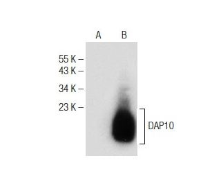
: sc-374196. Western blot analysis of DAP10 expression in non-transfected: sc-117752 (A) and human DAP10 transfected: sc-116977 (B) 293T whole cell lysates.")
 Alexa Fluor® 790: sc-374196 AF790. Direct near-infrared western blot analysis of DAP10 expression in HEL 92.1.7 (A) and HL-60 (B) whole cell lysates. Blocked with UltraCruz® Blocking Reagent: sc-516214.")
DAP10 Antibody (H-3): sc-374196
- DAP10 Antibody (H-3) is a mouse monoclonal IgG2a κ, cited in 3 publications, provided at 200 µg/ml
- specific for an epitope mapping between amino acids 51-79 at the C-terminus of DAP10 of mouse origin
- Anti-DAP10 Antibody (H-3) is recommended for detection of DAP10 of mouse, rat and human origin by WB, IP, IF and ELISA
- Anti-DAP10 Antibody (H-3) is available conjugated to agarose for IP; HRP for WB, IHC(P) and ELISA; and to either phycoerythrin or FITC for IF, IHC(P) and FCM
- also available conjugated to Alexa Fluor® 488, Alexa Fluor® 546, Alexa Fluor® 594 or Alexa Fluor® 647 for WB (RGB), IF, IHC(P) and FCM, and for use with RGB fluorescent imaging systems, such as iBright™ FL1000, FluorChem™, Typhoon, Azure and other comparable systems
- also available conjugated to Alexa Fluor® 680 or Alexa Fluor® 790 for WB (NIR), IF and FCM; for use with Near-Infrared (NIR) detection systems, such as LI-COR®Odyssey®, iBright™ FL1000, FluorChem™, Typhoon, Azure and other comparable systems
- m-IgG Fc BP-HRP and m-IgGκ BP-HRP are the preferred secondary detection reagents for DAP10 Antibody (H-3) for WB applications. These reagents are now offered in bundles with DAP10 Antibody (H-3) (see ordering information below).

QUICK LINKS
SEE ALSO...
DAP10 Antibody (H-3) is a mouse monoclonal IgG2a kappa light chain antibody that detects DAP10 in mouse, rat, and human samples through applications such as western blotting (WB), immunoprecipitation (IP), immunofluorescence (IF), and enzyme-linked immunosorbent assay (ELISA). Anti-DAP10 antibody (H-3) is available in both non-conjugated and various conjugated forms, including agarose, horseradish peroxidase (HRP), phycoerythrin (PE), fluorescein isothiocyanate (FITC), and multiple Alexa Fluor® conjugates. DAP10 is a transmembrane type 1 protein predominantly expressed in hematopoietic cells, playing a crucial role in immune responses. DAP10′s structure includes a cytoplasmic domain that contains a binding site for the SH2 domain of the p85 subunit of phosphoinositide 3-kinase (PI 3-kinase), indicating DAP10′s function as a signal transducer that activates PI 3-kinase. This activation is vital for mediating cellular responses to stress and promoting the cytotoxic activity of Natural Killer cells and T-cells against tumors expressing ligands such as MICA and MICB. The DAP10-NKG2D complex, which is formed with DAP10′s physiological partner NKG2D, is essential for initiating immune responses against tumor cells, highlighting DAP10′s importance in cancer immunology. Additionally, DAP10′s glycosylation causes DAP10 to migrate slightly slower than expected on SDS-PAGE, which is a critical consideration for accurate protein analysis.
Alexa Fluor® is a trademark of Molecular Probes Inc., OR., USA
LI-COR® and Odyssey® are registered trademarks of LI-COR Biosciences
DAP10 Antibody (H-3) References:
- An activating immunoreceptor complex formed by NKG2D and DAP10. | Wu, J., et al. 1999. Science. 285: 730-2. PMID: 10426994
- Contrasting roles of DAP10 and KARAP/DAP12 signaling adaptors in activation of the RBL-2H3 leukemic mast cell line. | Anfossi, N., et al. 2003. Eur J Immunol. 33: 3514-22. PMID: 14635062
- Silencing human NKG2D, DAP10, and DAP12 reduces cytotoxicity of activated CD8+ T cells and NK cells. | Karimi, M., et al. 2005. J Immunol. 175: 7819-28. PMID: 16339517
- Cloning, sequencing, and cell surface expression pattern of bovine immunoreceptor NKG2D and adaptor molecules DAP10 and DAP12. | Fikri, Y., et al. 2007. Immunogenetics. 59: 653-9. PMID: 17530242
- HMBOX1 negatively regulates NK cell functions by suppressing the NKG2D/DAP10 signaling pathway. | Wu, L., et al. 2011. Cell Mol Immunol. 8: 433-40. PMID: 21706044
- A possible mechanism of impaired NK cytotoxicity in cancer patients: down-regulation of DAP10 by TGF-beta1. | Lee, JC., et al. 2011. Tumori. 97: 350-7. PMID: 21789015
- TGF-β1 down-regulation of NKG2D/DAP10 and 2B4/SAP expression on human NK cells contributes to HBV persistence. | Sun, C., et al. 2012. PLoS Pathog. 8: e1002594. PMID: 22438812
- A novel bispecific chimeric PD1-DAP10/NKG2D receptor augments NK92-cell therapy efficacy for human gastric cancer SGC-7901 cell. | Li, M., et al. 2020. Biochem Biophys Res Commun. 523: 745-752. PMID: 31952789
- T-cells expressing a chimeric-PD1-Dap10-CD3zeta receptor reduce tumour burden in multiple murine syngeneic models of solid cancer. | Parriott, G., et al. 2020. Immunology. 160: 280-294. PMID: 32144940
- TCR extracellular domain genetically linked to CD28, 2B4/41BB and DAP10/CD3ζ -engineered NK cells mediates antitumor effects. | Li, S., et al. 2023. Cancer Immunol Immunother. 72: 769-774. PMID: 35988132
Ordering Information
| Product Name | Catalog # | UNIT | Price | Qty | FAVORITES | |
DAP10 Antibody (H-3) | sc-374196 | 200 µg/ml | $322.00 | |||
DAP10 Antibody (H-3): m-IgG Fc BP-HRP Bundle | sc-529687 | 200 µg Ab; 10 µg BP | $361.00 | |||
DAP10 Antibody (H-3): m-IgGκ BP-HRP Bundle | sc-522657 | 200 µg Ab, 40 µg BP | $361.00 | |||
DAP10 Antibody (H-3) AC | sc-374196 AC | 500 µg/ml, 25% agarose | $424.00 | |||
DAP10 Antibody (H-3) HRP | sc-374196 HRP | 200 µg/ml | $322.00 | |||
DAP10 Antibody (H-3) FITC | sc-374196 FITC | 200 µg/ml | $336.00 | |||
DAP10 Antibody (H-3) PE | sc-374196 PE | 200 µg/ml | $349.00 | |||
DAP10 Antibody (H-3) Alexa Fluor® 488 | sc-374196 AF488 | 200 µg/ml | $364.00 | |||
DAP10 Antibody (H-3) Alexa Fluor® 546 | sc-374196 AF546 | 200 µg/ml | $364.00 | |||
DAP10 Antibody (H-3) Alexa Fluor® 594 | sc-374196 AF594 | 200 µg/ml | $364.00 | |||
DAP10 Antibody (H-3) Alexa Fluor® 647 | sc-374196 AF647 | 200 µg/ml | $364.00 | |||
DAP10 Antibody (H-3) Alexa Fluor® 680 | sc-374196 AF680 | 200 µg/ml | $364.00 | |||
DAP10 Antibody (H-3) Alexa Fluor® 790 | sc-374196 AF790 | 200 µg/ml | $364.00 | |||
DAP10 (H-3) Neutralizing Peptide | sc-374196 P | 100 µg/0.5 ml | $69.00 |