Date published: 2025-10-16

1-800-457-3801

SCBT Portrait Logo
Seach Input

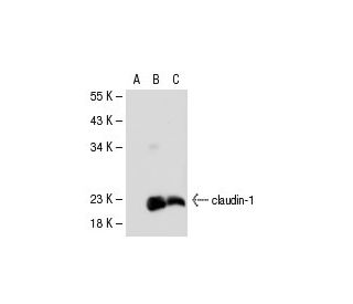
claudin-1 Antibody (XX7): sc-81796

5.0(10)
Write a reviewAsk a question

Datasheets
  • claudin-1 Antibody (XX7) is a mouse monoclonal IgG2a κ, cited in 25 publications, provided at 100 µg/ml
  • raised against recombinant claudin-1 of human origin
  • recommended for detection of claudin-1 of mouse, rat and human origin by WB, IP, IF, IHC(P) and ELISA
  • See claudin-1 (A-9): sc-166338 for claudin-1 antibody conjugates, including AC, HRP, FITC, PE, Alexa Fluor® 488, 594, 647, 680 and 790.
  • m-IgG Fc BP-HRP and m-IgG2a BP-HRP are the preferred secondary detection reagents for claudin-1 Antibody (XX7) for WB and IHC(P) applications. These reagents are now offered in bundles with claudin-1 Antibody (XX7) (see ordering information below).

QUICK LINKS

SEE ALSO...

Claudin-1 Antibody (XX7) is a mouse monoclonal IgG2a kappa light chain antibody raised against recombinant claudin-1 of human origin, detecting claudin-1 from human, mouse, and rat species through applications like western blotting (WB), immunoprecipitation (IP), immunofluorescence (IF), immunohistochemistry with paraffin-embedded sections (IHCP), and enzyme-linked immunosorbent assay (ELISA). Claudin-1, also known as CLDN1, is a critical component of the claudin family, involved in forming and maintaining tight junctions in epithelial and endothelial tissues. As a multi-pass membrane protein, claudin-1 is integral to cellular barriers that control paracellular transport, regulating ion permeability and molecule passage between cells. Tight junctions, which include claudin proteins along with occludins and junctional adhesion molecules, are essential for tissue integrity, especially in organs requiring strict barrier functions like the kidney and liver, where claudin-1 is most highly expressed. With four transmembrane domains and two extracellular loops, claudin-1 is structured to facilitate tight junction strand formation, which helps compartmentalize distinct tissue environments. In addition to its role in barrier function, altered claudin-1 expression is linked to various pathologies, with mutations in its gene leading to neonatal ichthyosis-sclerosing cholangitis syndrome, a rare autosomal recessive disorder. Anti-claudin-1 antibody (XX7) is designed to provide precise detection of this key protein across multiple species and applications.

For Research Use Only. Not Intended for Diagnostic or Therapeutic Use.

Ordering Information

Product NameCatalog #UNITPriceQtyFAVORITES

claudin-1 Antibody (XX7)

sc-81796
100 µg/ml
$333.00

claudin-1 Antibody (XX7): m-IgG Fc BP-HRP Bundle

sc-539665
100 µg Ab; 10 µg BP
$354.00

claudin-1 Antibody (XX7): m-IgG2a BP-HRP Bundle

sc-546745
100 µg Ab; 10 µg BP
$354.00

Can sc-81796: claudin-1 (XX7) monoclonal antibody be used to stain formalin-fixed, paraffin-embedded (FFPE) tissue sections?

Asked by: Cweed
Thank you for your question. Yes, sc-81796: claudin-1 (XX7) is recommended for use in IHC with paraffin-embedded sections. We recommend performing antigen retrieval with sodium citrate buffer (pH 6) and heat. The full protocol can be found here: https://www.scbt.com/scbt/resources/protocols/immunoperoxidase-staining
Answered by: Technical Support
Date published: 2017-03-24
  • y_2025, m_10, d_14, h_6CST
  • bvseo_bulk, prod_bvqa, vn_bulk_3.0.42
  • cp_1, bvpage1
  • co_hasquestionsanswers, tq_1
  • loc_en_US, sid_81796, prod, sort_[SortEntry(order=LAST_APPROVED_ANSWER_SUBMISSION_TIME, direction=DESCENDING)]
  • clientName_scbt
  • bvseo_sdk, java_sdk, bvseo-4.0.0
  • CLOUD, getContent, 177ms
  • QUESTIONS, PRODUCT
Rated 5 out of 5 by from westernで検出できました試供品で頂いたものを試しました。Western blottinのバンドが綺麗に出ましたので今後も使いたいと思います。
Date published: 2019-11-15
Rated 5 out of 5 by from Good for IF tissue sectionsI tested a sample of this on rat tissue cryosections, fixed with methanol and stained with this antibody overnight at 1:75 dilution. Works well with minimal to no background (depending on tissue).
Date published: 2018-02-20
Rated 5 out of 5 by from Produced publishable WB data in human and mouseseveral citations in the literature for Western Blotting on human K5+/K19− cells, myoepithelial progenitor cells and mouse wer back skin
Date published: 2017-01-25
Rated 5 out of 5 by from Strong band in WB in ECV304 whole cell lysateStrong band in WB in ECV304 whole cell lysate. -SCBT QC
Date published: 2015-05-04
Rated 5 out of 5 by from Produced great immunoperoxidase membraneProduced great immunoperoxidase membrane and cytoplasmic staining in formalin-fixed, paraffin-embedded human stomach tissue. -SCBT QC
Date published: 2015-04-25
Rated 5 out of 5 by from GoodGood, specific band in WB in human claudin-1 transfected 293T and SCC-4 whole cell lysates. -SCBT QC
Date published: 2015-04-17
Rated 5 out of 5 by from Published IHC(P) data for use in human epitheliumPublished IHC(P) data for use in human epithelium, adenoma, and adenocarcinoma sample. -SCBT Publication Review
Date published: 2015-03-20
Rated 5 out of 5 by from Strong band in WB in PRC/PRF/5 whole cell lysateStrong band in WB in PRC/PRF/5 whole cell lysate. -SCBT QC
Date published: 2015-03-18
  • y_2025, m_10, d_14, h_6
  • bvseo_bulk, prod_bvrr, vn_bulk_3.0.42
  • cp_1, bvpage1
  • co_hasreviews, tv_0, tr_10
  • loc_en_US, sid_81796, prod, sort_[SortEntry(order=SUBMISSION_TIME, direction=DESCENDING)]
  • clientName_scbt
  • bvseo_sdk, java_sdk, bvseo-4.0.0
  • CLOUD, getReviews, 8ms
  • REVIEWS, PRODUCT
claudin-1 Antibody (XX7) is rated 5.0 out of 5 by 10.
  • y_2025, m_10, d_14, h_6
  • bvseo_bulk, prod_bvrr, vn_bulk_3.0.42
  • cp_1, bvpage1
  • co_hasreviews, tv_0, tr_10
  • loc_en_US, sid_81796, prod, sort_[SortEntry(order=SUBMISSION_TIME, direction=DESCENDING)]
  • clientName_scbt
  • bvseo_sdk, java_sdk, bvseo-4.0.0
  • CLOUD, getAggregateRating, 117ms
  • REVIEWS, PRODUCT