")
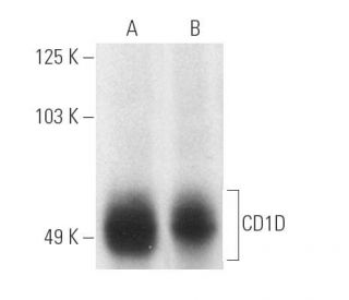
CD1D Antibody (C3D5): sc-19632
- CD1D Antibody (C3D5) is a mouse monoclonal IgG2a κ CD1D antibody, cited in 10 publications, provided at 200 µg/ml
- raised against the α 1 domain of CD1D of human origin
- CD1D Antibody (C3D5) is recommended for detection of an epitope in the α 1 domain of CD1D of human origin by WB, IP, IF and IHC(P)
- Anti-CD1D Antibody (C3D5) is available conjugated to agarose for IP; HRP for WB, IHC(P) and ELISA; and to either phycoerythrin or FITC for IF, IHC(P) and FCM
- also available conjugated to Alexa Fluor® 488, Alexa Fluor® 546, Alexa Fluor® 594 or Alexa Fluor® 647 for WB (RGB), IF, IHC(P) and FCM, and for use with RGB fluorescent imaging systems, such as iBright™ FL1000, FluorChem™, Typhoon, Azure and other comparable systems
- also available conjugated to Alexa Fluor® 680 or Alexa Fluor® 790 for WB (NIR), IF and FCM; for use with Near-Infrared (NIR) detection systems, such as LI-COR®Odyssey®, iBright™ FL1000, FluorChem™, Typhoon, Azure and other comparable systems
- m-IgGκ BP-HRP is the preferred secondary detection reagent for CD1D Antibody (C3D5) for WB and IHC(P) applications. This reagent is now offered in a bundle with CD1D Antibody (C3D5) (see ordering information below). For additional m-IgGκ BP conjugates see our complete list of Mouse IgG Binding Proteins.

QUICK LINKS
CD1D Antibody (C3D5) is a mouse monoclonal IgG2a kappa light chain antibody that detects the CD1D protein of human origin by western blotting (WB), immunoprecipitation (IP), immunofluorescence (IF), and immunohistochemistry. Anti-CD1D antibody (C3D5) is available in both non-conjugated and various conjugated forms, including agarose, horseradish peroxidase (HRP), phycoerythrin (PE), fluorescein isothiocyanate (FITC), and multiple Alexa Fluor® conjugates. The CD1 multigene family encodes five forms of the CD1 T cell surface glycoprotein in humans, which are CD1A, CD1B, CD1C, CD1D, and CD1E. CD1D plays a crucial role in the immune system by presenting lipid antigens to T lymphocytes, thereby influencing T cell activation and maturation. CD1D is primarily located on the surface of human intestinal epithelial cells and is known to exist in two forms: a β-2-Microglobulin-independent nonglycosylated form and a β-2-Microglobulin-dependent glycosylated form. CD1D expression on these cells facilitates the recognition of lipid antigens, which is essential for the immune response against various pathogens. Additionally, CD1D is associated with β-2-Microglobulin and is expressed on cortical thymocytes, Langerhans cells, a subset of B cells, and certain dendritic cells, highlighting its importance in the development and activation of CD1-restricted T cells. The human CD1D gene is located on chromosome 1q23.1 and encodes a 335 amino acid protein that is vital for normal T cell maturation, making CD1D (C3D5) monoclonal antibody an essential tool for researchers studying immune responses and T cell biology.
Alexa Fluor® is a trademark of Molecular Probes Inc., OR., USA
LI-COR® and Odyssey® are registered trademarks of LI-COR Biosciences
CD1D Antibody (C3D5) References:
- Human CD1d associates with prolyl-4-hydroxylase during its biosynthesis. | Kim, HS., et al. 2000. Mol Immunol. 37: 861-8. PMID: 11257307
- CD1d and natural T cells: how their properties jump-start the immune system. | Joyce, S. 2001. Cell Mol Life Sci. 58: 442-69. PMID: 11315191
- The identification of the beta 2-microglobulin binding antigen encoded by the human CD1D gene. | Bilsland, CA. and Milstein, C. 1991. Eur J Immunol. 21: 71-8. PMID: 1703966
- Isolation and characterization of a cDNA and gene coding for a fourth CD1 molecule. | Balk, SP., et al. 1989. Proc Natl Acad Sci U S A. 86: 252-6. PMID: 2463622
- Two classes of CD1 genes. | Calabi, F., et al. 1989. Eur J Immunol. 19: 285-92. PMID: 2467814
- Lipids regulate peripheral serotonin release via gut CD1d. | Luo, J., et al. 2023. Immunity. 56: 1533-1547.e7. PMID: 37354904
- Enhanced CD1d phosphatidylserine presentation using a single-domain antibody promotes immunomodulatory CD1d-TIM-3 interactions. | Lameris, R., et al. 2023. J Immunother Cancer. 11: PMID: 38040419
- CD1d protects against hepatocyte apoptosis in non-alcoholic steatohepatitis. | Lei, Z., et al. 2024. J Hepatol. 80: 194-208. PMID: 38438948
- The CD1 family: a third lineage of antigen-presenting molecules. | Porcelli, SA. 1995. Adv Immunol. 59: 1-98. PMID: 7484459
- Beta 2-microglobulin-independent MHC class Ib molecule expressed by human intestinal epithelium. | Balk, SP., et al. 1994. Science. 265: 259-62. PMID: 7517575
- Antigen presentation by CD1 and MHC-encoded class I-like molecules. | Melián, A., et al. 1996. Curr Opin Immunol. 8: 82-8. PMID: 8729450
- Analysis of the requirement for beta 2-microglobulin for expression and formation of human CD1 antigens. | Bauer, A., et al. 1997. Eur J Immunol. 27: 1366-73. PMID: 9209486
Ordering Information
| Product Name | Catalog # | UNIT | Price | Qty | FAVORITES | |
CD1D Antibody (C3D5) | sc-19632 | 200 µg/ml | $322.00 | |||
CD1D Antibody (C3D5): m-IgGκ BP-HRP Bundle | sc-520694 | 200 µg Ab, 40 µg BP | $361.00 | |||
CD1D Antibody (C3D5) AC | sc-19632 AC | 500 µg/ml, 25% agarose | $424.00 | |||
CD1D Antibody (C3D5) HRP | sc-19632 HRP | 200 µg/ml | $322.00 | |||
CD1D Antibody (C3D5) FITC | sc-19632 FITC | 200 µg/ml | $336.00 | |||
CD1D Antibody (C3D5) PE | sc-19632 PE | 200 µg/ml | $349.00 | |||
CD1D Antibody (C3D5) Alexa Fluor® 488 | sc-19632 AF488 | 200 µg/ml | $364.00 | |||
CD1D Antibody (C3D5) Alexa Fluor® 546 | sc-19632 AF546 | 200 µg/ml | $364.00 | |||
CD1D Antibody (C3D5) Alexa Fluor® 594 | sc-19632 AF594 | 200 µg/ml | $364.00 | |||
CD1D Antibody (C3D5) Alexa Fluor® 647 | sc-19632 AF647 | 200 µg/ml | $364.00 | |||
CD1D Antibody (C3D5) Alexa Fluor® 680 | sc-19632 AF680 | 200 µg/ml | $364.00 | |||
CD1D Antibody (C3D5) Alexa Fluor® 790 | sc-19632 AF790 | 200 µg/ml | $364.00 |