")
CD1C Anticuerpo (B-6): sc-390980
- CD1C Anticuerpo B-6 es un monoclonal de ratón IgG1 κ CD1C Anticuerpo, ver las 1 publicaciones, proporcionado como 200 µg/ml
- producido contra los amino ácidos 74-118 localizados en la región interna de CD1C de origen human
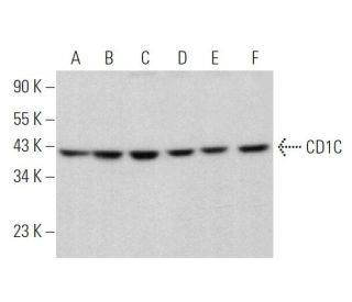
- CD1C Anticuerpo (B-6) es recomendado para detectar CD1C de mouse, rat y human origen, mediante WB, IP, IF y ELISA
- CD1C Anticuerpo (B-6) es disponible conjugado a agarosa para IP; HRP para WB, IHC(P) y ELISA; y tanto a phycoerythrin como a FITC para IF, IHC(P) y FCM
- también disponible conjugado a Alexa Fluor® 488, Alexa Fluor® 546, Alexa Fluor® 594 o Alexa Fluor® 647 para WB (RGB), IF, IHC (P) y FCM
- también disponible conjugado a Alexa Fluor® 680 o Alexa Fluor® 790 para WB (NIR), IF y FCM
- m-IgG Fc BP-HRP, 1 BP-HRP">m-IgG1 BP-HRP y m-IgGκ BP-HRP son los reactivos de detección secundarios preferidos para CD1C Anticuerpo (B-6) para aplicaciones WB. Estos reactivos se ofrecen ahora en paquetes con CD1C Anticuerpo (B-6)(véase la información de pedido más abajo).

El anticuerpo CD1C (B-6) es un anticuerpo monoclonal de ratón IgG1 κ CD1C (también designado como anticuerpo CD1C) que detecta la proteína CD1C de origen humano, de ratón y de rata mediante WB, IP, IF y ELISA. El anticuerpo CD1C (B-6) está disponible tanto en su forma no conjugada como en múltiples formas conjugadas, incluyendo agarosa, HRP, PE, FITC y múltiples conjugados de Alexa Fluor®. La familia multigénica CD1 codifica cinco formas de la glicoproteína de superficie de células T CD1 en humanos, designadas como CD1A, 1B, 1C, 1D y 1E. CD1, una proteína de membrana tipo 1, tiene similitud estructural con el antígeno de clase I del MHC y se ha demostrado que presenta antígenos lipídicos para el reconocimiento por linfocitos T. Los antígenos CD1 están asociados con β-2-Microglobulina y se expresan en timocitos corticales, células de Langerhans, un subconjunto de células B y algunas células dendríticas. Específicamente, CD1A es un marcador para la histiocitosis de células de Langerhans (LCH) y se encuentra en células interdigitantes. Los complejos de adaptadores de proteínas y los chaperones asociados a CD1 controlan el tráfico de CD1, y el desarrollo y activación de células T restringidas a CD1. La endocitosis constitutiva de las moléculas de CD1B y la clasificación diferencial de MHC de clase II de los lisosomas separan las moléculas presentadoras de antígenos peptídicos y lipídicos durante la maduración de las células dendríticas. CD1B también se expresa en células interdigitantes. Los genes humanos CD1 están todos estrechamente vinculados en un grupo que se mapea en el cromosoma 1q 22-23.
Alexa Fluor® es una marca registrada de Molecular Probes Inc., OR., USA
REIVEW LI-COR® y Odyssey® son marcas registradas de LI-COR Biosciences.
CD1C Anticuerpo (B-6) Referencias:
- Nuevos conocimientos sobre las vías de presentación de antígenos mediadas por CD1. | Sugita, M., et al. 2004. Curr Opin Immunol. 16: 90-5. PMID: 14734115
- CD1: presentación de antígenos y función de las células T. | Brigl, M. and Brenner, MB. 2004. Annu Rev Immunol. 22: 817-90. PMID: 15032598
- Estructura y expresión de los antígenos timocitarios humanos CD1a, CD1b y CD1c. | Martin, LH., et al. 1987. Proc Natl Acad Sci U S A. 84: 9189-93. PMID: 2447586
- La expresión de clones de ADNc que codifican los antígenos timocitarios CD1a, b, c demuestra una jerarquía de exclusión en los fibroblastos. | Aruffo, A. and Seed, B. 1989. J Immunol. 143: 1723-30. PMID: 2701945
- Clonación molecular de CD1a (T6), un marcador de células dendríticas epidérmicas humanas relacionado con moléculas MHC de clase I. | Longley, J., et al. 1989. J Invest Dermatol. 92: 628-31. PMID: 2784820
- La familia CD1: un tercer linaje de moléculas presentadoras de antígenos. | Porcelli, SA. 1995. Adv Immunol. 59: 1-98. PMID: 7484459
- Antígenos de superficie de poblaciones de timocitos humanos definidos por la expresión de CD3, CD4 y CD8: CD1a se expresa en los timocitos maduros pero no en las células T periféricas. | Sotzik, F., et al. 1993. Immunol Lett. 36: 101-6. PMID: 7688345
- Los CD1A-C de clase I no protegen a las células diana de la citólisis mediada por NK. | Storkus, WJ., et al. 1996. Cell Immunol. 167: 154-6. PMID: 8548840
- Presentación de antígenos por CD1 y moléculas de clase I codificadas por el CMH. | Melián, A., et al. 1996. Curr Opin Immunol. 8: 82-8. PMID: 8729450
- Análisis del requisito de beta 2-microglobulina para la expresión y formación de antígenos CD1 humanos. | Bauer, A., et al. 1997. Eur J Immunol. 27: 1366-73. PMID: 9209486
Información sobre pedidos
| Nombre del producto | Número de catálogo | UNIDAD | Precio | CANTIDAD | Favoritos | |
CD1C Anticuerpo (B-6) | sc-390980 | 200 µg/ml | $322.00 | |||
Paquete de CD1C (B-6): m-IgG Fc BP-HRP | sc-530324 | 200 µg Ab; 10 µg BP | $361.00 | |||
Paquete de CD1C (B-6): m-IgGκ BP-HRP | sc-523762 | 200 µg Ab, 40 µg BP | $361.00 | |||
Paquete de CD1C (B-6): m-IgG1 BP-HRP | sc-543990 | 200 µg Ab; 20 µg BP | $361.00 | |||
CD1C Anticuerpo (B-6) AC | sc-390980 AC | 500 µg/ml, 25% agarose | $424.00 | |||
CD1C Anticuerpo (B-6) HRP | sc-390980 HRP | 200 µg/ml | $322.00 | |||
CD1C Anticuerpo (B-6) FITC | sc-390980 FITC | 200 µg/ml | $336.00 | |||
CD1C Anticuerpo (B-6) PE | sc-390980 PE | 200 µg/ml | $349.00 | |||
CD1C Anticuerpo (B-6) Alexa Fluor® 488 | sc-390980 AF488 | 200 µg/ml | $364.00 | |||
CD1C Anticuerpo (B-6) Alexa Fluor® 546 | sc-390980 AF546 | 200 µg/ml | $364.00 | |||
CD1C Anticuerpo (B-6) Alexa Fluor® 594 | sc-390980 AF594 | 200 µg/ml | $364.00 | |||
CD1C Anticuerpo (B-6) Alexa Fluor® 647 | sc-390980 AF647 | 200 µg/ml | $364.00 | |||
CD1C Anticuerpo (B-6) Alexa Fluor® 680 | sc-390980 AF680 | 200 µg/ml | $364.00 | |||
CD1C Anticuerpo (B-6) Alexa Fluor® 790 | sc-390980 AF790 | 200 µg/ml | $364.00 |