")
Anticorps cathepsin S (E-3): sc-271619
- L'anticorps cathepsin S E-3 est un monoclonal IgG1 κ L'Anticorps cathepsin S, cité dans 35 publications, fourni en 200 µg/ml
- spécifique d'un épitope situé entre les acides aminés 302-331 au niveau C-terminus de cathepsin S d'origine human
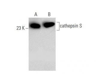
- L'Anticorps cathepsin S (E-3) est recommandé pour la détection de cathepsin S d'origine mouse, rat et human par WB, IP, IF, IHC(P) et ELISA
- Anti-L'Anticorps cathepsin S (E-3) est disponible conjugué à l'agarose pour IP; à l'HRP pour WB, IHC(P) et ELISA; et soit à la phycoerythrin ou FITC pour IF, IHC(P) et FCM
- aussi disponible conjugué à l'Alexa Fluor® 488, Alexa Fluor® 546, Alexa Fluor® 594 ou Alexa Fluor® 647 pour WB (RGB), IF, IHC(P) et FCM
- aussi disponible conjugué à l'Alexa Fluor® 680 ou Alexa Fluor® 790 pour WB (NIR), IF et FCM
- m-IgG Fc BP-HRP et m-IgG1 BP-HRP sont les réactifs de détection secondaire préférés pour cathepsin S Antibody (E-3) for WB and IHC(P) applications. Ces réactifs sont désormais proposés en lots avec cathepsin S Antibody (E-3)(voir les informations de commande ci-dessous).

ACCÈS RAPIDE AUX LIENS
VOIR ÉGALEMENT...
L'anticorps anti-cathepsine S (E-3) est un anticorps monoclonal IgG1 κ de souris contre la cathepsine S (également appelé anticorps CTSS) qui détecte la protéine cathepsine S de souris, de rat et d'origine humaine par WB, IP, IF, IHC(P) et ELISA. L'anticorps anti-cathepsine S (E-3) est disponible sous forme d'anticorps anti-cathepsine S non conjugué, ainsi que sous plusieurs formes conjuguées d'anticorps anti-cathepsine S, y compris agarose, HRP, PE, FITC et plusieurs conjugués Alexa Fluor®. La famille des enzymes protéolytiques cathepsines comprend plusieurs classes de protéases. La classe des protéases à cystéine comprend les cathepsines B, L, H, K, S et O. La classe des protéases à aspartyle est composée des cathepsines D et E. La cathepsine G fait partie de la classe des protéases à sérine. La plupart des cathepsines sont lysosomales et chacune d'entre elles est impliquée dans le métabolisme cellulaire, participant à divers événements tels que la biosynthèse des peptides et la dégradation des protéines. Il a été démontré que la cathepsine S est une cystéine protéinase élastinolytique présente dans les macrophages aveyronnais.
Alexa Fluor® est une marque déposée de Molecular Probes Inc., OR., USA
LI-COR® et Odyssey® sont marques déposées de LI-COR Biosciences
Anticorps cathepsin S (E-3) Références:
- Clonage moléculaire et expression de la cathepsine S du macrophage alvéolaire humain, une cystéine protéase élastinolytique. | Shi, GP., et al. 1992. J Biol Chem. 267: 7258-62. PMID: 1373132
- Organisation moléculaire du gène de la cathepsine D humaine. | Redecker, B., et al. 1991. DNA Cell Biol. 10: 423-31. PMID: 2069717
- La cathepsine S comme cible dans le cancer gastrique. | da Costa, AC., et al. 2020. Mol Clin Oncol. 12: 99-103. PMID: 31929878
- L'élafine inverse la fibrose intestinale en inhibant le récepteur 2 activé par la protéase de la cathepsine S. | Xie, Y., et al. 2022. Cell Mol Gastroenterol Hepatol. 14: 841-876. PMID: 35840034
- L'inhibition de la cathepsine S supprime le remodelage artériel pulmonaire associé au lupus érythémateux systémique expérimental. | Yen, TH., et al. 2022. Int J Mol Sci. 23: PMID: 36293172
- Clonage moléculaire et séquençage de l'ADNc de la cathepsine L de rat. | Ishidoh, K., et al. 1987. FEBS Lett. 223: 69-73. PMID: 3666143
- Clonage moléculaire et séquençage de l'ADNc de la cathepsine H de rat. Homologie dans les régions pro-peptidiques des protéinases à cystéine. | Ishidoh, K., et al. 1987. FEBS Lett. 226: 33-7. PMID: 3691815
- Isolement et séquençage de deux clones d'ADNc codant pour la cathepsine E de la rate de rat et analyse de l'activation de la procathepsine E purifiée. | Okamoto, K., et al. 1995. Arch Biochem Biophys. 322: 103-11. PMID: 7574663
- Clonage moléculaire de la cathepsine O humaine, une nouvelle endoprotéinase et homologue de l'OC2 du lapin. | Shi, GP., et al. 1995. FEBS Lett. 357: 129-34. PMID: 7805878
- La cathepsine B, une protéase à cystéine impliquée dans la progression métastatique, est également exprimée lors de la régression de la prostate et des glandes mammaires chez le rat. | Guenette, RS., et al. 1994. Eur J Biochem. 226: 311-21. PMID: 8001549
- Clonage moléculaire, localisation chromosomique et expression tissulaire spécifique du gène de la cathepsine G murine. | Heusel, JW., et al. 1993. Blood. 81: 1614-23. PMID: 8453108
- Cathepsine K de la souris: clonage de l'ADNc et expression prédominante du gène dans les ostéoclastes et dans certains chondrocytes hypertrophiés au cours du développement de la souris. | Rantakokko, J., et al. 1996. FEBS Lett. 393: 307-13. PMID: 8814310
Informations pour la commande
| Nom du produit | Ref. Catalogue | COND. | Prix HT | QTÉ | Favoris | |
Anticorps cathepsin S (E-3) | sc-271619 | 200 µg/ml | $322.00 | |||
cathepsin S (E-3): m-IgG Fc BP-HRP Kit | sc-527268 | 200 µg Ab; 10 µg BP | $361.00 | |||
cathepsin S (E-3): m-IgG1 BP-HRP Kit | sc-532641 | 200 µg Ab; 20 µg BP | $361.00 | |||
Anticorps cathepsin S (E-3) AC | sc-271619 AC | 500 µg/ml, 25% agarose | $424.00 | |||
Anticorps cathepsin S (E-3) HRP | sc-271619 HRP | 200 µg/ml | $322.00 | |||
Anticorps cathepsin S (E-3) FITC | sc-271619 FITC | 200 µg/ml | $336.00 | |||
Anticorps cathepsin S (E-3) PE | sc-271619 PE | 200 µg/ml | $349.00 | |||
Anticorps cathepsin S (E-3) Alexa Fluor® 488 | sc-271619 AF488 | 200 µg/ml | $364.00 | |||
Anticorps cathepsin S (E-3) Alexa Fluor® 546 | sc-271619 AF546 | 200 µg/ml | $364.00 | |||
Anticorps cathepsin S (E-3) Alexa Fluor® 594 | sc-271619 AF594 | 200 µg/ml | $364.00 | |||
Anticorps cathepsin S (E-3) Alexa Fluor® 647 | sc-271619 AF647 | 200 µg/ml | $364.00 | |||
Anticorps cathepsin S (E-3) Alexa Fluor® 680 | sc-271619 AF680 | 200 µg/ml | $364.00 | |||
Anticorps cathepsin S (E-3) Alexa Fluor® 790 | sc-271619 AF790 | 200 µg/ml | $364.00 | |||
cathepsin S (E-3) peptide neutralisant | sc-271619 P | 100 µg/0.5 ml | $69.00 |