")
ALG-2 Antibody (H-11): sc-376950
- ALG-2 Antibody (H-11) is a mouse monoclonal IgG1 κ ALG-2 antibody, cited in 1 publications, provided at 200 µg/ml
- raised against amino acids 1-191 representing full length ALG-2 of human origin
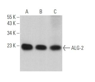
- ALG-2 Antibody (H-11) is recommended for detection of ALG-2 of mouse, rat and human origin by WB, IP, IF and ELISA
- Anti-ALG-2 Antibody (H-11) is available conjugated to agarose for IP; HRP for WB, IHC(P) and ELISA; and to either phycoerythrin or FITC for IF, IHC(P) and FCM
- also available conjugated to Alexa Fluor® 488, Alexa Fluor® 546, Alexa Fluor® 594 or Alexa Fluor® 647 for WB (RGB), IF, IHC(P) and FCM, and for use with RGB fluorescent imaging systems, such as iBright™ FL1000, FluorChem™, Typhoon, Azure and other comparable systems
- also available conjugated to Alexa Fluor® 680 or Alexa Fluor® 790 for WB (NIR), IF and FCM; for use with Near-Infrared (NIR) detection systems, such as LI-COR®Odyssey®, iBright™ FL1000, FluorChem™, Typhoon, Azure and other comparable systems
- m-IgG Fc BP-HRP and m-IgG1 BP-HRP are the preferred secondary detection reagents for ALG-2 Antibody (H-11) for WB applications. These reagents are now offered in bundles with ALG-2 Antibody (H-11) (see ordering information below).

QUICK LINKS
ALG-2 Antibody (H-11) is a mouse monoclonal IgG1 kappa light chain antibody that detects ALG-2 protein of mouse, rat, and human origin by western blotting (WB), immunoprecipitation (IP), immunofluorescence (IF), and enzyme-linked immunosorbent assay (ELISA). ALG-2 (H-11) antibody is available in both non-conjugated and various conjugated forms, including agarose, horseradish peroxidase (HRP), phycoerythrin (PE), fluorescein isothiocyanate (FITC), and multiple Alexa Fluor® conjugates, providing versatility for different experimental needs. ALG-2 protein, known for its role in apoptosis, is crucial for regulating cell death pathways, particularly in response to increased intracellular calcium concentrations. ALG-2 is predominantly expressed in the brain, with heightened levels observed in ischemic conditions, highlighting potential involvement in neurodegenerative processes. Structurally, ALG-2 contains five EF-hand-like motifs, which are essential for calcium-binding capability, allowing participation in the late stages of apoptosis where various death signals converge. ALG-2 interaction with other proteins, such as calpain small subunits, sorcin, and grancalcin, underscores importance in cellular signaling and apoptosis regulation, making ALG-2 a valuable target for research in cell death mechanisms and related diseases.
Alexa Fluor® is a trademark of Molecular Probes Inc., OR., USA
LI-COR® and Odyssey® are registered trademarks of LI-COR Biosciences
ALG-2 Antibody (H-11) References:
- Increased expression of apoptosis-linked gene 2 (ALG2) in the rat brain after temporary focal cerebral ischemia. | Li, W., et al. 2000. Neuroscience. 96: 161-8. PMID: 10683420
- Two forms of the apoptosis-linked protein ALG-2 with different Ca(2+) affinities and target recognition. | Tarabykina, S., et al. 2000. J Biol Chem. 275: 10514-8. PMID: 10744743
- The role of Ca2+ in cell killing. | Nicotera, P., et al. 1990. Chem Res Toxicol. 3: 484-94. PMID: 2103319
- Calcium-dependent killing of immature thymocytes by stimulation via the CD3/T cell receptor complex. | McConkey, DJ., et al. 1989. J Immunol. 143: 1801-6. PMID: 2528581
- Glucocorticoids activate a suicide process in thymocytes through an elevation of cytosolic Ca2+ concentration. | McConkey, DJ., et al. 1989. Arch Biochem Biophys. 269: 365-70. PMID: 2537063
- Interfering with apoptosis: Ca(2+)-binding protein ALG-2 and Alzheimer's disease gene ALG-3. | Vito, P., et al. 1996. Science. 271: 521-5. PMID: 8560270
- Functional cloning of genes involved in T-cell receptor-induced programmed cell death. | D'Adamio, L., et al. 1997. Semin Immunol. 9: 17-23. PMID: 9106304
- A growing family of the Ca2+-binding proteins with five EF-hand motifs. | Maki, M., et al. 1997. Biochem J. 328 (Pt 2): 718-20. PMID: 9441591
- Localization of mRNA for the apoptosis-linked gene ALG-2 in young and aged rat brain. | Venn, MK. and Conway, EL. 1998. Neuroreport. 9: 1981-5. PMID: 9674578
Ordering Information
| Product Name | Catalog # | UNIT | Price | Qty | FAVORITES | |
ALG-2 Antibody (H-11) | sc-376950 | 200 µg/ml | $322.00 | |||
ALG-2 Antibody (H-11): m-IgG Fc BP-HRP Bundle | sc-527557 | 200 µg Ab; 10 µg BP | $361.00 | |||
ALG-2 Antibody (H-11): m-IgG1 BP-HRP Bundle | sc-532930 | 200 µg Ab; 20 µg BP | $361.00 | |||
ALG-2 Antibody (H-11) AC | sc-376950 AC | 500 µg/ml, 25% agarose | $424.00 | |||
ALG-2 Antibody (H-11) HRP | sc-376950 HRP | 200 µg/ml | $322.00 | |||
ALG-2 Antibody (H-11) FITC | sc-376950 FITC | 200 µg/ml | $336.00 | |||
ALG-2 Antibody (H-11) PE | sc-376950 PE | 200 µg/ml | $349.00 | |||
ALG-2 Antibody (H-11) Alexa Fluor® 488 | sc-376950 AF488 | 200 µg/ml | $364.00 | |||
ALG-2 Antibody (H-11) Alexa Fluor® 546 | sc-376950 AF546 | 200 µg/ml | $364.00 | |||
ALG-2 Antibody (H-11) Alexa Fluor® 594 | sc-376950 AF594 | 200 µg/ml | $364.00 | |||
ALG-2 Antibody (H-11) Alexa Fluor® 647 | sc-376950 AF647 | 200 µg/ml | $364.00 | |||
ALG-2 Antibody (H-11) Alexa Fluor® 680 | sc-376950 AF680 | 200 µg/ml | $364.00 | |||
ALG-2 Antibody (H-11) Alexa Fluor® 790 | sc-376950 AF790 | 200 µg/ml | $364.00 |