")
Anticorps Akt1 (F-8): sc-271149
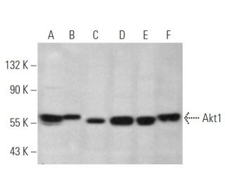
- L'Anticorps Akt1 (F-8) est un monoclonal de souris IgG1 κ, cité dans 14 publications, fourni en 200 µg/ml
- opposé à un peptide correspondant à C-terminus (h) de Akt1 d'origine human.
- recommandé pour la détection de Akt1 d'origine mouse, rat et human par WB, IP, IF, IHC(P) et ELISA
- Voir Akt1 (B-1): sc-5298 pour d'autres anticorps conjugués Akt1, dont AC, HRP, FITC, PE, Alexa Fluor® 488, 594, 647, 680 et 790.
- m-IgG Fc BP-HRP et m-IgG1 BP-HRP sont les réactifs de détection secondaire préférés pour Akt1 Antibody (F-8) for WB and IHC(P) applications. Ces réactifs sont désormais proposés en lots avec Akt1 Antibody (F-8)(voir les informations de commande ci-dessous).

ACCÈS RAPIDE AUX LIENS
La famille des sérine/thréonine kinases Akt contient plusieurs membres, dont Akt1 (également désigné PKB ou RacPK), Akt2 (également désigné PKBβ ou RacPK-βb) et Akt 3 (également désigné PKBγ ou proto-oncogène viral thyome 3), qui présentent une homologie de séquence avec les familles de protéines kinases A et C et sont codés par le proto-oncogène c-Akt. Tous les membres de la famille Akt possèdent un domaine d'homologie pleckstrine. Akt1 et Akt2 sont activés par la stimulation du PDGF. L'activation dépend des résidus Tyr 740 et 751 du PDGFR-β, qui lient la sous-unité du complexe phosphatidylinositol 3-kinase (PI 3-kinase). L'activation de l'Akt1 par l'insuline ou le facteur de croissance de l'insuline-1 (IGF-1) entraîne la phosphorylation des résidus Thr 308 et Ser 473. La phosphorylation de ces deux résidus est importante pour générer un niveau élevé d'activité de l'Akt1. La phosphorylation du Thr 308 ne dépend pas de la phosphorylation du Ser 473 in vivo. Ainsi, les protéines Akt sont phosphorylées et activées dans les cellules stimulées par l'insuline/IGF-1 par une (des) kinase(s) en amont. L'activation d'Akt1 et d'Akt2 est inhibée par l'inhibiteur de PI kinase wortmannin, ce qui suggère que la protéine émet un signal en aval des PI kinases.
Informations pour la commande
| Nom du produit | Ref. Catalogue | COND. | Prix HT | QTÉ | Favoris | |
Anticorps Akt1 (F-8) | sc-271149 | 200 µg/ml | $322.00 | |||
Akt1 (F-8): m-IgG Fc BP-HRP Kit | sc-540213 | 200 µg Ab; 10 µg BP | $361.00 | |||
Akt1 (F-8): m-IgG1 BP-HRP Kit | sc-541975 | 200 µg Ab; 20 µg BP | $361.00 | |||
Akt1 (F-8) peptide neutralisant | sc-271149 P | 100 µg/0.5 ml | $69.00 |