")
TBK1 Antibody (A-6): sc-398366
- TBK1 Antibody (A-6) is a mouse monoclonal IgG1 κ TBK1 antibody, cited in 12 publications, provided at 200 µg/ml
- raised against amino acids 355-729 mapping at the C-terminus of TBK1 of mouse origin
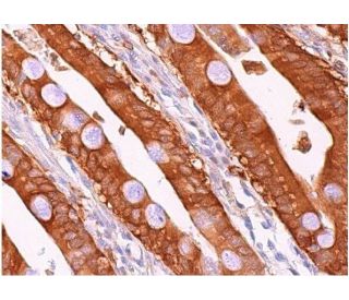
- TBK1 Antibody (A-6) is recommended for detection of TBK1 of mouse, rat and human origin by WB, IP, IF, IHC(P) and ELISA
- Anti-TBK1 Antibody (A-6) is available conjugated to agarose for IP; HRP for WB, IHC(P) and ELISA; and to either phycoerythrin or FITC for IF, IHC(P) and FCM
- also available conjugated to Alexa Fluor® 488, Alexa Fluor® 546, Alexa Fluor® 594 or Alexa Fluor® 647 for WB (RGB), IF, IHC(P) and FCM, and for use with RGB fluorescent imaging systems, such as iBright™ FL1000, FluorChem™, Typhoon, Azure and other comparable systems
- also available conjugated to Alexa Fluor® 680 or Alexa Fluor® 790 for WB (NIR), IF and FCM; for use with Near-Infrared (NIR) detection systems, such as LI-COR®Odyssey®, iBright™ FL1000, FluorChem™, Typhoon, Azure and other comparable systems
- m-IgG Fc BP-HRP, m-IgG1 BP-HRP and m-IgGκ BP-HRP are the preferred secondary detection reagents for TBK1 Antibody (A-6) for WB and IHC(P) applications. These reagents are now offered in bundles with TBK1 Antibody (A-6) (see ordering information below).

QUICK LINKS
SEE ALSO...
TBK1 Antibody (A-6) is a mouse monoclonal IgG1 kappa light chain antibody that detects TBK1 protein of mouse, rat, and human origin by western blotting (WB), immunoprecipitation (IP), immunofluorescence (IF), immunohistochemistry with paraffin-embedded sections (IHCP), and enzyme-linked immunosorbent assay (ELISA). anti-TBK1 antibody (A-6) is available in both non-conjugated and various conjugated forms, including agarose, horseradish peroxidase (HRP), phycoerythrin (PE), fluorescein isothiocyanate (FITC), and multiple Alexa Fluor® conjugates. TBK1 plays a crucial role in regulating immune response and inflammation, particularly through involvement in activating transcription factor NF-kB. This activation is significant because NF-kB drives expression of various pro-inflammatory cytokines and survival factors, which are vital for immune system response to pathogens and stress. TBK1 interacts with other proteins in NF-kB signaling pathway, including TRAF2 and TANK, facilitating phosphorylation and degradation of inhibitory protein IkB, which allows NF-kB to translocate to nucleus and activate target genes. TBK1′s ability to modulate this pathway underscores its importance in both innate immunity and pathogenesis of various inflammatory diseases, making TBK1 (A-6) monoclonal antibody a valuable tool for researchers studying these processes.
Alexa Fluor® is a trademark of Molecular Probes Inc., OR., USA
LI-COR® and Odyssey® are registered trademarks of LI-COR Biosciences
TBK1 Antibody (A-6) References:
- STING specifies IRF3 phosphorylation by TBK1 in the cytosolic DNA signaling pathway. | Tanaka, Y. and Chen, ZJ. 2012. Sci Signal. 5: ra20. PMID: 22394562
- TRIM23 mediates virus-induced autophagy via activation of TBK1. | Sparrer, KMJ., et al. 2017. Nat Microbiol. 2: 1543-1557. PMID: 28871090
- Spatiotemporal Control of ULK1 Activation by NDP52 and TBK1 during Selective Autophagy. | Vargas, JNS., et al. 2019. Mol Cell. 74: 347-362.e6. PMID: 30853401
- TBK1 recruitment to STING activates both IRF3 and NF-κB that mediate immune defense against tumors and viral infections. | Yum, S., et al. 2021. Proc Natl Acad Sci U S A. 118: PMID: 33785602
- Tiliroside targets TBK1 to induce ferroptosis and sensitize hepatocellular carcinoma to sorafenib. | Yang, C., et al. 2023. Phytomedicine. 111: 154668. PMID: 36657316
- VANGL2 inhibits antiviral IFN-I signaling by targeting TBK1 for autophagic degradation. | Hu, Z., et al. 2023. Sci Adv. 9: eadg2339. PMID: 37352355
- ATG4B antagonizes antiviral immunity by GABARAP-directed autophagic degradation of TBK1. | Xie, W., et al. 2023. Autophagy. 19: 2853-2868. PMID: 37434364
- Optineurin provides a mitophagy contact site for TBK1 activation. | Yamano, K., et al. 2024. EMBO J. 43: 754-779. PMID: 38287189
- A TBK1 variant causes autophagolysosomal and motoneuron pathology without neuroinflammation in mice. | Brenner, D., et al. 2024. J Exp Med. 221: PMID: 38517332
- CDK4/6 Alters TBK1 Phosphorylation to Inhibit the STING Signaling Pathway in Prostate Cancer. | Li, W., et al. 2024. Cancer Res. 84: 2588-2606. PMID: 38861362
Ordering Information
| Product Name | Catalog # | UNIT | Price | Qty | FAVORITES | |
TBK1 Antibody (A-6) | sc-398366 | 200 µg/ml | $322.00 | |||
TBK1 Antibody (A-6): m-IgG Fc BP-HRP Bundle | sc-530775 | 200 µg Ab; 10 µg BP | $361.00 | |||
TBK1 Antibody (A-6): m-IgGκ BP-HRP Bundle | sc-524312 | 200 µg Ab, 40 µg BP | $361.00 | |||
TBK1 Antibody (A-6): m-IgG1 BP-HRP Bundle | sc-544247 | 200 µg Ab; 20 µg BP | $361.00 | |||
TBK1 Antibody (A-6) AC | sc-398366 AC | 500 µg/ml, 25% agarose | $424.00 | |||
TBK1 Antibody (A-6) HRP | sc-398366 HRP | 200 µg/ml | $322.00 | |||
TBK1 Antibody (A-6) FITC | sc-398366 FITC | 200 µg/ml | $336.00 | |||
TBK1 Antibody (A-6) PE | sc-398366 PE | 200 µg/ml | $349.00 | |||
TBK1 Antibody (A-6) Alexa Fluor® 488 | sc-398366 AF488 | 200 µg/ml | $364.00 | |||
TBK1 Antibody (A-6) Alexa Fluor® 546 | sc-398366 AF546 | 200 µg/ml | $364.00 | |||
TBK1 Antibody (A-6) Alexa Fluor® 594 | sc-398366 AF594 | 200 µg/ml | $364.00 | |||
TBK1 Antibody (A-6) Alexa Fluor® 647 | sc-398366 AF647 | 200 µg/ml | $364.00 | |||
TBK1 Antibody (A-6) Alexa Fluor® 680 | sc-398366 AF680 | 200 µg/ml | $364.00 | |||
TBK1 Antibody (A-6) Alexa Fluor® 790 | sc-398366 AF790 | 200 µg/ml | $364.00 |