Date published: 2025-9-9

001 800-1338-3838

SCBT Portrait Logo
Seach Input

CD81 항체 (5A6): sc-23962

4.1(14)
리뷰 쓰기질문하기

데이터 시트
  • CD81 항체(5A6) 는 마우스 monoclonal IgG1κ, 127간행물에 인용, 이며 200 µg/ml으로 제공합니다.
  • 안구 흑색종 세포주(V+B2)에 대해 항원화
  • 안티-CD81 항체 (5A6)는 WB, IP, IF and FCM으로 mouse, rat and human유래의 CD81 를 감지하는 데에 추천한다.
  • 항-CD81 항체(5A6)는 IP용 agarose와 결합되어 이용 가능하며, WB, IHC(P), ELISA용 HRP와 결합되어 이용 가능하며, IF, IHC(P), FCM용 phycoerythrin 또는 FITC와 결합되어 이용 가능합니다.
  • WB (RGB), IF, IHC(P) 와FCM, RGB fluorescent imaging systems, such as iBright™ FL1000, FluorChem™, Typhoon, Azure and other comparable systems에 사용가능한 Alexa Fluor® 488, Alexa Fluor® 546, Alexa Fluor® 594 or Alexa Fluor® 647결합제품도 있습니다.
  • WB (NIR), IF와FCM,Near-Infrared (NIR) detection systems, such as LI-COR®Odyssey®, iBright™ FL1000, FluorChem™, Typhoon, Azure and other comparable systems에 사용가능한 Alexa Fluor® 680 or Alexa Fluor® 790 결합제품도 있습니다.
  • CD81 (5A6): sc-23962무료10 µg 샘플을 신청하려면 기술지원팀 (또는 로컬 디스트리뷰터)에 연락주세요.
  • m-IgG Fc BP-HRP1 BP-HRP">m-IgG1 BP-HRP 는 CD81 항체 (5A6) for WB applications. 이 시약은 이제 CD81 항체 (5A6)와 함께 번들로 제공됩니다(아래 주문 정보 참조).

빠른 링크

더보기

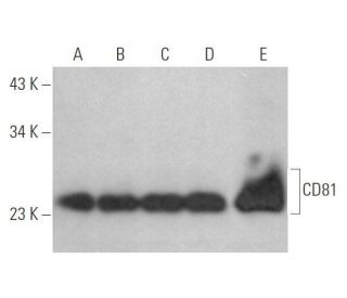
CD81 항체(5A6)는 4개의 막 관통 도메인을 특징으로 하는 테트라스파닌 계열의 두드러진 구성원인 TAPA-1로도 알려진 CD81 단백질을 표적으로 삼도록 특별히 설계된 마우스 단일 클론 IgG1 카파 경쇄 항체입니다. CD81은 조혈, 신경외배엽, 중간엽 기원의 세포에서 광범위하게 발현되며, 세포 성장, 신호 전달, 막 소구체의 조직화에 중요한 역할을 합니다. CD81은 팔미토일화 및 당화반응을 포함한 필수적인 번역 후 변형을 겪으며, 이는 적절한 국소화, 안정성, 그리고 CD37, CD53, CD19, CD21, MHC class II 항원과 같은 다른 막 단백질과의 상호작용에 매우 중요합니다. 항-CD81 항체(5A6)는 OCI-LY8 B 림프종 세포주를 사용하여 개발되었으며, 웨스턴 블랏팅(WB), 면역침전법(IP), 면역형광법(IF), 유세포분석법(FCM) 등의 응용을 통해 마우스, 랫트, 인간 종에서 CD81을 효과적으로 검출할 수 있습니다. 호스래디시 퍼옥시다아제(HRP), 피코에리틴(PE), 플루오레세인 이소티오시아네이트(FITC), 다양한 알렉사 플루오르® 접합체 등 접합체 형태와 비접합체 형태로 제공되는 CD81 단일 클론 항체(5A6)는 다양한 실험적 요구에 맞는 다양한 옵션을 제공합니다. 연구자들은 CD81을 표적으로 삼아 면역 세포 활성화, 바이러스 침입 메커니즘, 세포 기능을 관장하는 복잡한 신호 전달 경로와 같은 중요한 과정을 조사할 수 있습니다. CD81의 번역 후 변형은 암 전이 및 면역 장애와 같은 병리학적 조건에서 CD81의 역할을 이해하는 데 특히 중요하므로, 항-CD81 항체(5A6)는 기초 및 응용 연구에 필수적인 도구입니다.

연구용으로만 사용가능합니다. 진단이나 치료용으로 사용불가합니다.

Alexa Fluor®는 미국 오리건주 Molecular Probes Inc.의 상표입니다.

LI-COR®와 Odyssey®는 LI-COR Biosciences의 등록 상표입니다.

주문정보

제품명카탈로그 번호 단위가격수량관심품목

CD81 항체 (5A6)

sc-23962
200 µg/ml
$316.00

CD81 (5A6): m-IgG Fc BP-HRP 번들

sc-526643
200 µg Ab; 10 µg BP
$354.00

CD81 (5A6): m-IgG1 BP-HRP 번들

sc-532016
200 µg Ab; 20 µg BP
$354.00

CD81 항체 (5A6) AC

sc-23962 AC
500 µg/ml, 25% agarose
$416.00

CD81 항체 (5A6) HRP

sc-23962 HRP
200 µg/ml
$316.00

CD81 항체 (5A6) FITC

sc-23962 FITC
200 µg/ml
$330.00

CD81 항체 (5A6) PE

sc-23962 PE
200 µg/ml
$343.00

CD81 항체 (5A6) Alexa Fluor® 488

sc-23962 AF488
200 µg/ml
$357.00

CD81 항체 (5A6) Alexa Fluor® 546

sc-23962 AF546
200 µg/ml
$357.00

CD81 항체 (5A6) Alexa Fluor® 594

sc-23962 AF594
200 µg/ml
$357.00

CD81 항체 (5A6) Alexa Fluor® 647

sc-23962 AF647
200 µg/ml
$357.00

CD81 항체 (5A6) Alexa Fluor® 680

sc-23962 AF680
200 µg/ml
$357.00

CD81 항체 (5A6) Alexa Fluor® 790

sc-23962 AF790
200 µg/ml
$357.00

What is the isoelectric point for CD-81 antibody?

Asked by: Sumita
Thank you for your question. We do not characterize the isoelectric point of our antibodies.
Answered by: BlakeJ
Date published: 2025-07-23

Hello, I have used the CD81 antibody for western blot at 1:1500 in 5% MILK on exosomes from human plasma. Do you know why I get two bands, one at 23 kDa and another one at 48 kDa? Many thanks Juliana

Asked by: Juliana Redondo
Thank you for your question. The expected molecular weight for CD81 is ~ 22-26 kDa. We have not tested this antibody specifically in human plasma, and have not observed a higher molecular weight signal at 48 kDa in the cell lines tested. CD81 has been reported to dimerize (PMID: 20375010) and that may be a consideration. We would also recommend including a secondary antibody control, where you omit the primary antibody from the incubation, to confirm if the higher molecular weight band might be due to non-specific binding of the secondary antibody alone.
Answered by: Technical Support
Date published: 2017-10-16

Hi, I have used this antibody at 1:100 concentration on a western blot (10% SDS-PAGE gel) loading some exosomal protein. Could you explain please why I get not only the 26 kDa band, but also a band at about 50 kDa? Thank you.

Asked by: VivSal
Thank you for your question. We have not tested this antibody on exosomal protein, or observed a 50 kDa band in Western blot. We would recommend including a secondary antibody control (omit primary antibody) to identify if the 50 kDa band may be a result of non-specific binding of the secondary antibody alone. It could also be your secondary antibody binding to heavy chain IgG in your samples. Below is a link to our recommended protocol for Western blotting. Please contact our Technical Service Department if for additional support with troubleshooting the antibody. https://www.scbt.com/scbt/resources/protocols/western-immuno-blotting
Answered by: Technical Support
Date published: 2017-10-16

I have tried using this antibody on a western blot (after running an SDS-PAGE gel) with a homemade K562 lysate as control and did not get a signal at 1:100 dilution. I tried it 3 times previously on other cell lines at 1:200 w/no success. Any suggestions?

Asked by: Tali
Thank you for your question. If you have no signal, we would recommend verifying that your anti-mouse secondary is working well. We would also recommend including a loading control antibody (e.g., Actin or GAPDH) to ensure that your transfer conditions are good, as well as your signal detection conditions (e.g., chemiluminescence or other). We have successfully used this antibody for WB at a dilution of 1:200, loading 50 µg of K-562 whole cell lysate. A link to our complete recommended protocol is provided below. If you continue to have negative results, please contact our Technical Service Department so we can provide further troubleshooting support. Santa Cruz Biotechnology does warranty our antibodies for one year from the date of purchase. If we are unable to successfully troubleshoot your application we will send you a replacement antibody free of charge, or issue a credit or refund if that is not possible https://www.scbt.com/scbt/resources/protocols/western-immuno-blotting .
Answered by: Technical Support
Date published: 2017-04-26

What concentration would you recommend using SC-20048 at, to detect a protein in paraffin-embedded human tissue? We have used 1:1000 successfully for western blots. Please can you reply at your earliest convenience.

Asked by: Deborah Goberdhan
Thank you for your question! We recommend a starting dilution of 1:50 for IHC on paraffin embedded tissue sections. This is a starting dilution, so it is possible that you can use it much more diluted! We also recommend performing an antigen retrieval step, ideally sodium citrate with heat. You can find our recommended protocol here: https://www.scbt.com/scbt/resources/protocols/immunoperoxidase-staining If you have any further questions, please contact our European Technical Service team. You can reach them by phone at Toll Free: +49 6221 4503 0, by email at: europe@scbt.com or by live chat directly on our website, www.scbt.com
Answered by: Tech Service
Date published: 2017-02-22

1, What concentration do you recommend using SC-23962 on paraffin embedded human tissue sections? We have this antibody, so would like to try. ; 2. Do you think that SC-23962 FITC would work better and at what conc. for same application, ie IHC(P)?

Asked by: Deborah Goberdhan
Thank you for your question! We recommend a starting dilution of 1:50 for IHC on paraffin embedded tissue sections. We do recommend performing an antigen retrieval step. You can find our recommended protocol here: https://www.scbt.com/scbt/resources/protocols/immunoperoxidase-staining You can use the FITC conjugated form, although we do not generally use FITC conjugated antibodies for IHC in house, so I do not have test results for this. If you wish to try it, I would stick with the 1:50 starting dilution as well. If you have any further questions, please contact our European Technical Service team. You can reach them by phone at Toll Free: +49 6221 4503 0, by email at: europe@scbt.com or by live chat directly on our website, www.scbt.com
Answered by: Tech Service
Date published: 2017-02-22

Can I use this antibody for staining of mouse tissue, since it is a mouse antibody?

Asked by: Anna99
Yes. CD81 Antibody (5A6): sc-23962 is available conjugated to either phycoerythrin (sc-23962 PE) or fluoroscein (sc-23962 FITC), 200 µg/ml, for IF, IHC(P) and FCM.
Answered by: Tech Service 5
Date published: 2017-01-05
  • y_2025, m_9, d_7, h_4CST
  • bvseo_bulk, prod_bvqa, vn_bulk_3.0.42
  • cp_1, bvpage1
  • co_hasquestionsanswers, tq_7
  • loc_ko_KR, sid_23962, prod, sort_[SortEntry(order=LAST_APPROVED_ANSWER_SUBMISSION_TIME, direction=DESCENDING)]
  • clientName_scbt
  • bvseo_sdk, java_sdk, bvseo-4.0.0
  • CLOUD, getContent, 144ms
  • QUESTIONS, PRODUCT
Rated 4 out of 5 by from Great antibodyI request a sample of the antibody and it worked well!
Date published: 2023-07-31
Rated 4 out of 5 by from buon funzionamentoHo effettuato un solo esperimento per verificare la reattività nel cane e ha funzionato
Date published: 2023-07-14
Rated 1 out of 5 by from WBでバンドが検出できませんでした。エクソソームサンプルでCD81のバンドが全く検出できませんでした。同じサンプルで他のCD81抗体を使うとCD81を検出できたのでサンプルは問題ないと考えています。
Date published: 2021-09-09
Rated 2 out of 5 by from No band in Western blotI cannot see any band in Western blot after a nice transfer (all MW completely transfered; blotting using 1:200 of this antibody and 2nd antibody sc-516102 at 1:1000) of exosomes isolated by ultracentrifugation and further separated by density centrifugation. Perhaps I have low concentration of exosomes, but I expected to get at least a faint band.
Date published: 2021-05-25
Rated 5 out of 5 by from Works well in western blotThe antibody detected CD81 in Mesenchymal stem cells at the expected molecular size.
Date published: 2020-09-18
Rated 5 out of 5 by from Tested on HFFF by FACSThis antibody was tested on HFFF cells by FACS and shown a very strong signal, even at dilution 1:100
Date published: 2018-11-19
Rated 5 out of 5 by from Excellent antibody for ICC stainingThis antibody is gorgeous for ICC staining, please see the attached confocal image. Highly recommended. I tried 1:50 dilution for the first time, and you may further dilute it to 1:100 or even 1:200 to decrease the background a little bit, and it should work even better. I feel the overall quality of SCBT antibodies is getting much better,
Date published: 2018-11-08
Rated 5 out of 5 by from likeMembrane localization observed by Immunofluorescence staining of methanol-fixed embedded HeLa cells, also good for staining.
Date published: 2017-05-07
  • y_2025, m_9, d_7, h_4
  • bvseo_bulk, prod_bvrr, vn_bulk_3.0.42
  • cp_1, bvpage1
  • co_hasreviews, tv_0, tr_14
  • loc_ko_KR, sid_23962, prod, sort_[SortEntry(order=SUBMISSION_TIME, direction=DESCENDING)]
  • clientName_scbt
  • bvseo_sdk, java_sdk, bvseo-4.0.0
  • CLOUD, getReviews, 15ms
  • REVIEWS, PRODUCT
CD81 항체 (5A6) is rated 4.1 out of 5 by 14.
  • y_2025, m_9, d_7, h_4
  • bvseo_bulk, prod_bvrr, vn_bulk_3.0.42
  • cp_1, bvpage1
  • co_hasreviews, tv_0, tr_14
  • loc_ko_KR, sid_23962, prod, sort_[SortEntry(order=SUBMISSION_TIME, direction=DESCENDING)]
  • clientName_scbt
  • bvseo_sdk, java_sdk, bvseo-4.0.0
  • CLOUD, getAggregateRating, 111ms
  • REVIEWS, PRODUCT