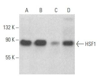
Date published: 2025-9-7

00800 4573 8000

SCBT Portrait Logo
Seach Input

Anticorps HSF1 (E-4): sc-17757

4.5(19)
Écrire une critiquePoser une question

Fiches techniques
  • L'anticorps HSF1 E-4 est un monoclonal IgG1 κ L'Anticorps HSF1, cité dans 51 publications, fourni en 200 µg/ml
  • dirigé contre les acides aminés 219-529 de heat shock transcription factor 1 (HSF1) d'origine human
  • L'Anticorps HSF1 (E-4) est recommandé pour la détection de HSF1 d'origine mouse, rat et human par WB, IP, IF, IHC(P) et ELISA
  • Anti-L'Anticorps HSF1 (E-4) est disponible conjugué à l'agarose pour IP; à l'HRP pour WB, IHC(P) et ELISA; et soit à la phycoerythrin ou FITC pour IF, IHC(P) et FCM
  • aussi disponible conjugué à l'Alexa Fluor® 488, Alexa Fluor® 546, Alexa Fluor® 594 ou Alexa Fluor® 647 pour WB (RGB), IF, IHC(P) et FCM
  • aussi disponible conjugué à l'Alexa Fluor® 680 ou Alexa Fluor® 790 pour WB (NIR), IF et FCM
  • Réactif TransCruz pour une utilisation en Gel Supershift et ChIP (sc-17757 X, 200 µg/0.1 ml)
  • Contactez notre Service Technique (ou votre Distributeur local) pour plus d'informations pour recevoir un échantillon GRATUIT de 10 µg de HSF1 (E-4): sc-17757.
  • m-IgGκ BP-HRP est le réactif secondaire recommandé pour la détection de l'anticorps HSF1 (E-4) pour des applications de WB and IHC(P). Ce réactif est maintenant proposé en kit avec l'anticorps HSF1 (E-4) (voir Informations pour la commande ci-dessous). Pour d'autres protéines de liaison m-IgGκ, veuillez vous référer à la liste complète des protéines de liaison IgG de souris.

ACCÈS RAPIDE AUX LIENS

VOIR ÉGALEMENT...

L'anticorps HSF1 (E-4) est un anticorps monoclonal IgG1 kappa à chaîne légère de souris qui détecte HSF1 d'origine humaine, murine et rat par western blotting (WB), immunoprécipitation (IP), immunofluorescence (IF), immunohistochimie (IHC) et enzyme-linked immunosorbent assay (ELISA). L'anticorps monoclonal HSF1 (E-4) est disponible sous forme non conjuguée et sous diverses formes conjuguées, notamment agarose, peroxydase de raifort, phycoérythrine, isothiocyanate de fluorescéine et plusieurs conjugués Alexa Fluor®. HSF1 joue un rôle crucial dans la réponse cellulaire au stress, notamment thermique et chimique, en régulant l'expression des protéines de choc thermique qui aident à protéger les cellules contre les dommages. Cette régulation est vitale pour le maintien de l'homéostasie cellulaire et la promotion de la survie des cellules dans des conditions défavorables. HSF1 est généralement présent sous forme de monomère, mais lorsqu'il est activé, il forme des trimères qui se déplacent vers le noyau, où HSF1 se lie aux éléments de choc thermique dans les régions promotrices des gènes cibles, facilitant ainsi la transcription. HSF1 est régulé à la hausse par les œstrogènes, qui renforcent son activité au niveau de l'ARNm et des protéines, ce qui souligne son importance dans les réponses cellulaires au stress et ses implications potentielles dans les maladies liées au stress et à l'inflammation.

Pour la Recherche Uniquement. Non destiné à un usage diagnostique ou thérapeutique.

Alexa Fluor® est une marque déposée de Molecular Probes Inc., OR., USA

LI-COR® et Odyssey® sont marques déposées de LI-COR Biosciences

Anticorps HSF1 (E-4) Références:

  1. Activation transcriptionnelle des gènes de choc thermique chez les eucaryotes.  |  Tanguay, RM. 1988. Biochem Cell Biol. 66: 584-93. PMID: 3048332
  2. Rôle de la protéine HSF1 dans la transformation maligne.  |  Šimončík, O., et al. 2018. Klin Onkol. 31: 55-62. PMID: 31023025
  3. Rôle de HSF1 et du réseau de chaperons dans le microenvironnement tumoral.  |  Grunberg, N., et al. 2020. Adv Exp Med Biol. 1243: 101-111. PMID: 32297214
  4. HSF1 est impliqué dans la réponse immunothérapeutique en régulant l'expression de PD-L1 médiée par APOJ/STAT3 dans le carcinome hépatocellulaire.  |  Cheng, H., et al. 2023. Cancer Biol Ther. 24: 1-9. PMID: 36482717
  5. Le facteur de choc thermique 1 (HSF1) potentialise spécifiquement la transcription médiée par c-MYC indépendamment de la réponse canonique au choc thermique.  |  Xu, M., et al. 2023. Cell Rep. 42: 112557. PMID: 37224019
  6. Les réponses transcriptionnelles des cellules cancéreuses aux stimuli induisant un choc thermique impliquent l'amplification de la liaison robuste de HSF1.  |  Dastidar, SG., et al. 2023. Nat Commun. 14: 7420. PMID: 37973875
  7. Expression dépendante des œstrogènes du facteur de transcription du choc thermique: implications pour la synthèse utérine des protéines de choc thermique.  |  Yang, X., et al. 1995. J Steroid Biochem Mol Biol. 52: 415-9. PMID: 7748806
  8. Détermination de la stœchiométrie du facteur 2 de transcription du choc thermique dans les complexes d'ADN en boucle à l'aide de la microscopie à force de balayage.  |  Wyman, C., et al. 1995. EMBO J. 14: 117-23. PMID: 7828583
  9. Fonction et régulation du facteur de choc thermique 2 au cours de l'embryogenèse de la souris.  |  Rallu, M., et al. 1997. Proc Natl Acad Sci U S A. 94: 2392-7. PMID: 9122205
  10. Réponse au choc thermique et dégradation des protéines: régulation de HSF2 par la voie ubiquitine-protéasome.  |  Mathew, A., et al. 1998. Mol Cell Biol. 18: 5091-8. PMID: 9710593
  11. L'inhibition du protéasome entraîne l'activation de tous les membres de la famille des facteurs de choc thermique.  |  Kawazoe, Y., et al. 1998. Eur J Biochem. 255: 356-62. PMID: 9716376
  12. La glycogène synthase kinase 3beta et la kinase régulée par le signal extracellulaire inactivent le facteur de transcription du choc thermique 1 en facilitant la disparition des granules transcriptionnellement actifs après le choc thermique.  |  He, B., et al. 1998. Mol Cell Biol. 18: 6624-33. PMID: 9774677

Informations pour la commande

Nom du produitRef. CatalogueCOND.Prix HTQTÉFavoris

Anticorps HSF1 (E-4)

sc-17757
200 µg/ml
$316.00

HSF1 (E-4): m-IgGκ BP-HRP Kit

sc-551063
200 µg Ab; 40 µg BP
$354.00

Anticorps HSF1 (E-4) X

sc-17757 X
200 µg/0.1 ml
$316.00

Anticorps HSF1 (E-4) AC

sc-17757 AC
500 µg/ml, 25% agarose
$416.00

Anticorps HSF1 (E-4) HRP

sc-17757 HRP
200 µg/ml
$316.00

Anticorps HSF1 (E-4) FITC

sc-17757 FITC
200 µg/ml
$330.00

Anticorps HSF1 (E-4) PE

sc-17757 PE
200 µg/ml
$343.00

Anticorps HSF1 (E-4) Alexa Fluor® 488

sc-17757 AF488
200 µg/ml
$357.00

Anticorps HSF1 (E-4) Alexa Fluor® 546

sc-17757 AF546
200 µg/ml
$357.00

Anticorps HSF1 (E-4) Alexa Fluor® 594

sc-17757 AF594
200 µg/ml
$357.00

Anticorps HSF1 (E-4) Alexa Fluor® 647

sc-17757 AF647
200 µg/ml
$357.00

Anticorps HSF1 (E-4) Alexa Fluor® 680

sc-17757 AF680
200 µg/ml
$357.00

Anticorps HSF1 (E-4) Alexa Fluor® 790

sc-17757 AF790
200 µg/ml
$357.00

Hello, I am a research technician at the University of Bristol, UK. I am interested in your HSF1 (E-4) antibody. Do you have any unconjugated version of this antibody? Can I have a free sample to try it to my cell line. Many thanks.

Posée par: Andriana
Thank you for your question. Please contact Technical Service for information on how to obtain a sample.
Répondue par: SCBT Heidelberg Support
Date de publication: 2018-12-14

I used 3ug of HSF1 (E-4) antibody for doing ChIP, but did not get much enrichment. Is there any recommended concentration of HSF1 antibody for ChIP that I should have used?

Posée par: S_G_
Thank you for your inquiry. For the IP step in ChIP we recommend using 100–500 µg supernatant and 0.1-1 µl TransCruz reagent (0.2–2 µg antibody). The complete protocol for ChIP can be found in below link. If you have any further questions, please call our Technical Service team toll-free at (800) 457-3801 option 2, e-mail <scbt@scbt.com>, or use the live chat on our website. Best of luck with your research! https://www.scbt.com/scbt/resources/protocols/chromatin-immunoprecipitation-assays
Répondue par: Technical Support 14
Date de publication: 2017-07-10

What is the recommended secondary reagent to be used with this mouse monoclonal primary?

Posée par: DefinitelyNotMatt
Thank you for your question. We recommend using one our exclusive Mouse IgG Binding Proteins as a secondary detection reagent. A complete list of available binding proteins is available on our website here: https://www.scbt.com/scbt/browse/support-products-mouse-igg-binding-proteins/_/N-ecrety Please contact Technical Service if you have further questions concerning this product.
Répondue par: Technical Support 12
Date de publication: 2017-01-13

Can I use this mouse monoclonal antibody for immunohistochemical staining of mouse tissue? Or even double or triple staining on the same samples?

Posée par: DefinitelyNotMatt
Thank you for your question. Yes. We recommend using a directly conjugated mouse monoclonal primary antibody. This antibody is currently available conjugated to phycoerythrin (sc-17757 PE), fluorescein (sc-17757 FITC), Alexa Fluor® 488 (sc-17757 AF488) or Alexa Fluor® 647 (sc-17757 AF647). Please contact Technical Service if you have further questions concerning this product.
Répondue par: Technical Support 12
Date de publication: 2017-01-13
  • y_2025, m_8, d_27, h_5CST
  • bvseo_bulk, prod_bvqa, vn_bulk_3.0.42
  • cp_1, bvpage1
  • co_hasquestionsanswers, tq_4
  • loc_fr_FR, sid_17757, prod, sort_[SortEntry(order=LAST_APPROVED_ANSWER_SUBMISSION_TIME, direction=DESCENDING)]
  • clientName_scbt
  • bvseo_sdk, java_sdk, bvseo-4.0.0
  • CLOUD, getContent, 103ms
  • QUESTIONS, PRODUCT
Rated 5 de 5 de par works goodworks good for my sample. cell line over-expressed with HSF1. 1:1000, clear background. but not sure for detecting endogenous level.
Date de publication: 2022-01-25
Rated 1 de 5 de par Not good for WB using human whole cell lysate.This antibody is not good for WB using human K562 whole cell lysate at 1:500 dilution.
Date de publication: 2021-08-05
Rated 2 de 5 de par Not working for my samplesI’ve tried this antibody on sub cellular fractionated PBMCs Lysates at 1:100 dilution but the signal was very low
Date de publication: 2020-09-02
Rated 3 de 5 de par works okayfaint signals were observed - samples were human cells
Date de publication: 2017-11-21
Rated 5 de 5 de par specifc signaWe got specific bands at 90kda with diution 1:800 by WB.HepG2 cell lysates 60ug.I don't recommend loading than 80ug, we have tried this amout ,it cause very high background,also I recommend use the secondary antibody sc-516102.
Date de publication: 2017-09-16
Rated 5 de 5 de par 功能很出色the antibody is very good,economical and practical
Date de publication: 2017-09-12
Rated 4 de 5 de par acceptable antibody for immunofluorescenceDilution 1:200 for IF on PFA fixed U2OS cells gives week signal (compared to polyclonal rabbit Ab). 1:50 should be tested.
Date de publication: 2017-08-08
Rated 5 de 5 de par specific binding target proteinI use HSF1antibody in our WB assay.The antibody is very full bind the target protein,specific and 1:500 is suitable but for the loading amount I recommend controled at 40 to 70 ug lysates.If more than this range,it may affect the specificity and singal intensity.
Date de publication: 2017-07-15
  • y_2025, m_8, d_27, h_5
  • bvseo_bulk, prod_bvrr, vn_bulk_3.0.42
  • cp_1, bvpage1
  • co_hasreviews, tv_0, tr_19
  • loc_fr_FR, sid_17757, prod, sort_[SortEntry(order=SUBMISSION_TIME, direction=DESCENDING)]
  • clientName_scbt
  • bvseo_sdk, java_sdk, bvseo-4.0.0
  • CLOUD, getReviews, 18ms
  • REVIEWS, PRODUCT
Anticorps HSF1 (E-4) est évalué 4.5 de 5 de 19.
  • y_2025, m_8, d_27, h_5
  • bvseo_bulk, prod_bvrr, vn_bulk_3.0.42
  • cp_1, bvpage1
  • co_hasreviews, tv_0, tr_19
  • loc_fr_FR, sid_17757, prod, sort_[SortEntry(order=SUBMISSION_TIME, direction=DESCENDING)]
  • clientName_scbt
  • bvseo_sdk, java_sdk, bvseo-4.0.0
  • CLOUD, getAggregateRating, 112ms
  • REVIEWS, PRODUCT