")
V-ATPase A1 항체 (E-8): sc-374475
- V-ATPase A1 항체 E-8 는 마우스 monoclonal IgG2b κ V-ATPase A1 항체, 10간행물에 인용, 이며 200 µg/ml으로 제공합니다.
- human의 V-ATPase A1의 세포외 도메인 내의 71-210 아미노산에 대한 항체
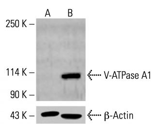
- V-ATPase A1 항체 (E-8)는 WB, IP, IF, IHC(P) and ELISA으로 mouse, rat and human유래의 V-ATPase A1 를 감지하는 데에 추천한다.; 이외에, equine, bovine, porcine and canine등 species와 반응할수 있습니다
- 항-V-ATPase A1 항체(E-8)는 IP용 agarose와 결합되어 이용 가능하며, WB, IHC(P), ELISA용 HRP와 결합되어 이용 가능하며, IF, IHC(P), FCM용 phycoerythrin 또는 FITC와 결합되어 이용 가능합니다.
- WB (RGB), IF, IHC(P) 와FCM, RGB fluorescent imaging systems, such as iBright™ FL1000, FluorChem™, Typhoon, Azure and other comparable systems에 사용가능한 Alexa Fluor® 488, Alexa Fluor® 546, Alexa Fluor® 594 or Alexa Fluor® 647결합제품도 있습니다.
- WB (NIR), IF와FCM,Near-Infrared (NIR) detection systems, such as LI-COR®Odyssey®, iBright™ FL1000, FluorChem™, Typhoon, Azure and other comparable systems에 사용가능한 Alexa Fluor® 680 or Alexa Fluor® 790 결합제품도 있습니다.
- m-IgGκ BP-HRP는 V-ATPase A1 항체 (E-8) WB 및 IHC(P) 애플리케이션용입니다. 이 시약은 현재 V-ATPase A1 항체 (E-8)(아래 주문 정보 참조)와 함께 번들로 제공됩니다. 추가 m-IgGκ BP 접합체는 마우스 IgG 결합 단백질 전체 목록을 참조하세요.

빠른 링크
V-ATPase A1 항체(E-8)는 IgG2b κ 마우스 단일 클론 V-ATPase A1 항체(ATP6V0A1 항체, VPP1 항체, ATPase H+ 운반 V0 서브유닛 A1 항체, ATP6N1A 항체, ATP6N1 항체, Stv1 항체, VPP1 항체, Vph1 항체, A1 항체로도 지정됨)입니다, 마우스, 쥐 및 인간 기원의 V-ATPase A1 단백질을 검출하는 ATPase H+ 운반 리소좀 비촉매 보조 단백질 1(110/116kD) 항체, V-ATPase 116 KDa 서브유닛 A1 항체 또는 V-ATPase 116 KDa 항체) WB, IP, IF, IHC(P) 및 ELISA로 측정합니다. V-ATPase A1 항체(E-8)는 비접합 항-V-ATPase A1 항체 형태와 아가로스, HRP, PE, FITC 및 여러 Alexa Fluor® 접합체를 포함한 여러 접합 형태의 항-V-ATPase A1 항체 형태로 제공됩니다. 진공 양성자 펌프의 서브유닛은 두 가지 다른 이소폼을 가진 V-ATPase입니다. I형 동형에는 18염기쌍 인서트가 포함되어 있으며 뇌에서 발현되는 반면, 잘린 II형 동형은 폐, 신장, 비장 등 더 광범위하게 발현됩니다. 진공 양성자 펌프의 서브유닛은 클라트린으로 코팅된 소포에 위치하며 파골세포에서도 발견됩니다. 이는 두 가지 기본 도메인, 즉 30% 이상의 하전 잔기가 있는 친수성 아미노 말단과 최소 6개의 막 통과 영역을 포함하는 소수성 카르복시 말단으로 구성됩니다. 양성자 펌프는 세포질 소단위에 의한 ATP 가수분해를 펌프의 막 내 구성 요소에 의한 양성자 전위에 결합하는 기능을 합니다. 파골세포 특이적 진공 양성자 ATPase 서브유닛의 비활성화는 골경화성 돌연변이 마우스에서 파골세포의 정단막에 효소가 부족하여 이들 세포의 재흡수 기능을 방해하고 골형성 표현형을 유발하는 원인이 됩니다. 이 서브유닛은 소포체 막에서 후기 소포체 마커인 Rab7과 공동 위치하며, 후기 소포체의 선택적 팽창에 의한 소포체 형성에 필수적입니다.
Alexa Fluor®는 미국 오리건주 Molecular Probes Inc.의 상표입니다.
LI-COR®와 Odyssey®는 LI-COR Biosciences의 등록 상표입니다.
V-ATPase A1 항체 (E-8) 참고문헌:
- 인간 파골세포 특이 116-kDa V-ATPase 서브유닛의 마우스 상동체를 암호화하는 유전자는 골경화성(oc/oc) 돌연변이체에서 결손이 있습니다. | Scimeca, JC., et al. 2000. Bone. 26: 207-13. PMID: 10709991
- 살리실아미드 A는 바포로마이신 A1과는 다른 메커니즘을 통해 V-ATPase의 V0 부문을 억제합니다. | Xie, XS., et al. 2004. J Biol Chem. 279: 19755-63. PMID: 14998996
- 클라트린 코팅 소포/시냅스 소포 양성자 펌프의 116-kDa 폴리펩타이드 구조. | Perin, MS., et al. 1991. J Biol Chem. 266: 3877-81. PMID: 1704894
- 바필로마이신 A1은 V-ATPase 의존성 산성화와 Ca-P60A/SERCA 의존성 오토파고좀-리소좀 융합을 억제함으로써 자가포식 작용을 방해합니다. | Mauvezin, C. and Neufeld, TP. 2015. Autophagy. 11: 1437-8. PMID: 26156798
- 망막 색소 상피 세포에서 βA3/A1-크리스탈린에 의한 V-ATPase 조절. | Valapala, M., et al. 2016. Adv Exp Med Biol. 854: 779-84. PMID: 26427489
- ABCA1(ATP 결합 카세트 단백질 A1)-매개 콜레스테롤 유출에 필요한 V-ATPase(진공 ATPase) 활성. | Lorkowski, SW., et al. 2018. Arterioscler Thromb Vasc Biol. 38: 2615-2625. PMID: 30354238
- 양성자 펌핑 V-ATPase 억제제인 바포로마이신 A1은 Rab7 리소좀의 위치에 영향을 미치고 파골세포 분비 리소좀의 전방 이동을 억제합니다. | Matsumoto, N. and Nakanishi-Matsui, M. 2019. Biochem Biophys Res Commun. 510: 421-426. PMID: 30717974
- 바필로마이신 A1에 의한 V-ATPase 억제의 분자적 기초. | Wang, R., et al. 2021. Nat Commun. 12: 1782. PMID: 33741963
- V-ATPase a1 서브유닛과 다른 이소폼의 질병 관련 돌연변이에 대한 구조적, 기능적 이해. | Indrawinata, K., et al. 2023. Front Mol Neurosci. 16: 1135015. PMID: 37465367
- 대체 mRNA 스플라이싱은 진공 양성자 펌프의 116-kDa 폴리펩타이드의 조직 특이적 이소형을 생성합니다. | Peng, SB., et al. 1994. J Biol Chem. 269: 17262-6. PMID: 8006034
- 헬리코박터 파일로리균에 의해 유도된 세포 내 액포에는 액포 ATPase 양성자 펌프가 존재합니다. | Papini, E., et al. 1996. J Med Microbiol. 45: 84-9. PMID: 8683556
- 진공 양성자 전위 ATPase의 116-kDa 하위 단위의 동소체 식별 및 재구성. | Peng, SB., et al. 1999. J Biol Chem. 274: 2549-55. PMID: 9891027
주문정보
| 제품명 | 카탈로그 번호 | 단위 | 가격 | 수량 | 관심품목 | |
V-ATPase A1 항체 (E-8) | sc-374475 | 200 µg/ml | $322.00 | |||
V-ATPase A1 (E-8): m-IgGκ BP-HRP 번들 | sc-522752 | 200 µg Ab, 40 µg BP | $361.00 | |||
V-ATPase A1 항체 (E-8) AC | sc-374475 AC | 500 µg/ml, 25% agarose | $424.00 | |||
V-ATPase A1 항체 (E-8) HRP | sc-374475 HRP | 200 µg/ml | $322.00 | |||
V-ATPase A1 항체 (E-8) FITC | sc-374475 FITC | 200 µg/ml | $336.00 | |||
V-ATPase A1 항체 (E-8) PE | sc-374475 PE | 200 µg/ml | $349.00 | |||
V-ATPase A1 항체 (E-8) Alexa Fluor® 488 | sc-374475 AF488 | 200 µg/ml | $364.00 | |||
V-ATPase A1 항체 (E-8) Alexa Fluor® 546 | sc-374475 AF546 | 200 µg/ml | $364.00 | |||
V-ATPase A1 항체 (E-8) Alexa Fluor® 594 | sc-374475 AF594 | 200 µg/ml | $364.00 | |||
V-ATPase A1 항체 (E-8) Alexa Fluor® 647 | sc-374475 AF647 | 200 µg/ml | $364.00 | |||
V-ATPase A1 항체 (E-8) Alexa Fluor® 680 | sc-374475 AF680 | 200 µg/ml | $364.00 | |||
V-ATPase A1 항체 (E-8) Alexa Fluor® 790 | sc-374475 AF790 | 200 µg/ml | $364.00 |