
Date published: 2026-10-20

1-800-457-3801

SCBT Portrait Logo
Seach Input

SOCS-3 Antikörper (SO1): sc-51699

4.8(9)
Produkt bewertenBitte stellen Sie eine Frage
Datenblätter
  • SOCS-3 Antikörper SO1 ist ein monoklonales IgG2b aus der Maus, verwendet in 18 wissenschaftlichen Veröffentlichungen, in einer Menge von 100 µg/ml
  • gegen ein gereinigtes natives SOC3-Protein voller Länge human
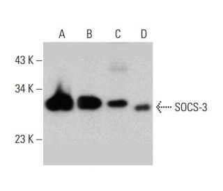
  • Empfohlen für die Detektion von SOCS-3 aus der Spezies mouse, rat und human per WB, IP und ELISA
  • 2b BP-HRP">m-IgG2b BP-HRP ist das bevorzugte sekundäre Nachweisreagenz für SOCS-3 Antikörper (SO1) für WB-Anwendungen. Dieses Reagenz wird jetzt in einem Paket mit SOCS-3 Antikörper (SO1) angeboten(siehe Bestellinformationen unten).

Gene Editing Promo Banner

Direktverknüpfungen

Siehe auch...

Der SOCS-3-Antikörper (SO1) ist ein monoklonaler Maus Ig2bSOCS-3-Antikörper, der SOCS-3 von maus-, ratten- und menschlicher Herkunft durch WB, IP und ELISA nachweist. Der SOCS-3-Antikörper (SO1) ist als nicht konjugierte Form des Anti-SOCS-3-Antikörpers erhältlich. Die SOCS-Genfamilie (Suppressor of Cytokine Signaling) besteht aus einer Gruppe von Proteinen, die die Signalübertragung von Zytokinen negativ regulieren. Die SOCS-Familienproteine enthalten ein zentrales SH2-Domänen- und ein carboxy-terminales Region, das als "SOCS-Box" bezeichnet wird. Die Gene SOCS-1 (auch SSI-1 und JAB genannt), SOCS-2 (auch SSI-2 und CIS2 genannt) und SOCS-3 (auch SSI-3 und CIS3 genannt) werden bekanntlich durch IL-6 und andere Zytokine hochreguliert. SOCS-4, SOCS-5, SOCS-6 und SOCS-7 wurden durch ihre Sequenzhomologie mit der SOCS-Box identifiziert. CIS (für cytokine-induzierte SH2-enthaltende Proteine) ist ebenfalls ein Mitglied der SOCS-Familie.

Ausschließlich für Forschungszwecke. Nicht für diagnostische oder therapeutische Zwecke bestimmt.

Alexa Fluor® ist ein Markenzeichen von Molecular Probes Inc., OR., USA

LI-COR® und Odyssey® sind Markenzeichen von LI-COR Biosciences

SOCS-3 Antikörper (SO1) Literaturhinweise:

  1. SOCS-3 induziert die Myoblastendifferenzierung.  |  Spangenburg, EE. 2005. J Biol Chem. 280: 10749-58. PMID: 15653674
  2. Methylierung von SOCS-3 und SOCS-1 bei der Karzinogenese des Barrett-Adenokarzinoms.  |  Tischoff, I., et al. 2007. Gut. 56: 1047-53. PMID: 17376806
  3. Die Rolle des SOCS-3-Proteins bei Leptinresistenz und Fettleibigkeit.  |  Lubis, AR., et al. 2008. Acta Med Indones. 40: 89-95. PMID: 18560028
  4. Expression und funktionelle Bedeutung von SOCS-1 und SOCS-3 in Astrozyten.  |  Qin, H., et al. 2008. J Immunol. 181: 3167-76. PMID: 18713987
  5. Die SOCS-1- und SOCS-3-Phosphorylierung ist für die Bcr-Abl-induzierte Tumorentstehung erforderlich.  |  Qiu, X., et al. 2012. Neoplasia. 14: 547-58. PMID: 22787435
  6. Expression von SOCS-1 und SOCS-3 im Myokard von Patienten mit plötzlichem Herztod.  |  Chen, L., et al. 2010. World J Emerg Med. 1: 99-103. PMID: 25214949
  7. Eine geringe Expression von SOCS-1 und SOCS-3 ist ein schlechter Prognoseindikator für Magenkrebspatienten.  |  Li, G., et al. 2015. J Cancer Res Clin Oncol. 141: 443-52. PMID: 25261987
  8. Bedeutung von SOCS-3 und SOCS-4 in vitro und mögliche mechanistische Zusammenhänge mit der Wundheilung.  |  Feng, Y., et al. 2017. Sci Rep. 7: 6715. PMID: 28751715
  9. Gewebespiegel von Suppressor of Cytokine Signaling-3 (SOCS-3) bei Mycosis fungoides.  |  Nada, HR., et al. 2023. Arch Dermatol Res. 315: 165-171. PMID: 35226171
  10. Auswirkungen von Valproinsäure auf die Genexpression von SOCS-1, SOCS-2, SOCS-3, SOCS-5, SOCS-6 und SOCS-7 und die Hemmung des Zellwachstums bei Dickdarmkarzinomen.  |  Sanaei, M., et al. 2022. Gastroenterol Hepatol Bed Bench. 15: 39-44. PMID: 35611249

Bestellinformation

ProduktKatalog #EINHEITPreisANZAHLFavoriten

SOCS-3 Antikörper (SO1)

sc-51699
100 µg/ml
$322.00

SOCS-3 (SO1): m-IgG2b BP-HRP Bundle

sc-548433
100 µg Ab; 10 µg BP
$361.00

Can we use this antibody for IF assay? If yes, what concentration do you suggest? If no, do you have SOCS3 antibodies for IF?

Gefragt von: as11
Thank you for your question. We have not yet tested this antibody for IF, or immunofluorescent staining, so this application is not recommended at this time. Unfortunately, we do not have any alternative antibodies against SOCS-3 that are recommended for IF at this time.
Beantwortet von: Technical Support
Veröffentlichungsdatum: 2017-07-27

For Western Blot, is it recommended to use denatured or non-denatured conditions with SOCS-3 (SO1): sc-51699 antibody?

Gefragt von: Dr Ninau Qelp
Thank you for your question. We recommend this antibody for use in denatured Western Blot conditions. It has not been validated for use in non-denatured conditions. Please contact our Technical Service Department for further details or inquiries.
Beantwortet von: Technical Support
Veröffentlichungsdatum: 2017-03-27
  • y_2026, m_10, d_20, h_5CST
  • bvseo_bulk, prod_bvqa, vn_bulk_3.0.48
  • cp_1, bvpage1
  • co_hasquestionsanswers, tq_2
  • loc_de_DE, sid_51699, prod, sort_[SortEntry(order=LAST_APPROVED_ANSWER_SUBMISSION_TIME, direction=DESCENDING)]
  • clientName_scbt
  • bvseo_sdk, java_sdk, bvseo-4.0.0
  • CLOUD, getContent, 103ms
  • QUESTIONS, PRODUCT
Rated 5 von 5 von janin aus good results in WBstrong and specific band in western blot wit mice liver lysates, dilution 1:1000
Veröffentlichungsdatum: 2018-09-05
Rated 5 von 5 von yanli aus 细胞免疫荧光刚试用了细胞免疫荧光,抗体说明书中没有表明可以用于此实验,但在293T细胞中试用结果很好,主要染色胞质。
Veröffentlichungsdatum: 2017-10-26
Rated 5 von 5 von nichaoying aus This antibody worked greatThis antibody worked great. I strongly recommend this antibody for detection of SOCS-3.
Veröffentlichungsdatum: 2017-10-23
Rated 4 von 5 von M__T_ aus Worked in WB using mouse/human cellsWorked in WB using mouse/human cells.
Veröffentlichungsdatum: 2015-09-15
Rated 5 von 5 von SM aus WB: human and mouse cell lysatesWB: human and mouse cell lysates. -SCBT Publication Review
Veröffentlichungsdatum: 2015-05-18
Rated 5 von 5 von JL aus Published western blot data using human bloodPublished western blot data using human blood samples. -SCBT Publication Review
Veröffentlichungsdatum: 2015-04-03
Rated 5 von 5 von FL aus Nice band in WB using HeLa cell lysatesNice band in WB using HeLa cell lysates. -SCBT QC
Veröffentlichungsdatum: 2015-01-21
Rated 4 von 5 von SH aus Positive western blot results in HeLaPositive western blot results in HeLa, IFN -treated HeLa, IL-6-treated HeLa and human PBL whole cell lysates. -SCBT QC
Veröffentlichungsdatum: 2015-01-11
  • y_2026, m_10, d_20, h_5
  • bvseo_bulk, prod_bvrr, vn_bulk_3.0.48
  • cp_1, bvpage1
  • co_hasreviews, tv_0, tr_9
  • loc_de_DE, sid_51699, prod, sort_[SortEntry(order=SUBMISSION_TIME, direction=DESCENDING)]
  • clientName_scbt
  • bvseo_sdk, java_sdk, bvseo-4.0.0
  • CLOUD, getReviews, 18ms
  • REVIEWS, PRODUCT
SOCS-3 Antikörper (SO1) wurde bewertet mit 4.8 von 5 von 9.
  • y_2026, m_10, d_20, h_5
  • bvseo_bulk, prod_bvrr, vn_bulk_3.0.48
  • cp_1, bvpage1
  • co_hasreviews, tv_0, tr_9
  • loc_de_DE, sid_51699, prod, sort_[SortEntry(order=SUBMISSION_TIME, direction=DESCENDING)]
  • clientName_scbt
  • bvseo_sdk, java_sdk, bvseo-4.0.0
  • CLOUD, getAggregateRating, 99ms
  • REVIEWS, PRODUCT