")
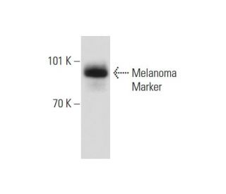
: sc-59305. Western blot analysis of Melanoma Marker expression in SK-MEL-28 whole cell lysate.")
 Alexa Fluor® 790: sc-59305 AF790. Direct near-infrared western blot analysis of Melanoma Marker expression in A-431 whole cell lysate. Blocked with UltraCruz® Blocking Reagent: sc-516214. Cruz Marker™ Molecular Weight Standards detected with Cruz Marker MW Tag-Alexa Fluor® 680: sc-516730.")
Melanoma Marker Antibody (HMB45): sc-59305
- Melanoma Marker Antibody (HMB45) is a mouse monoclonal IgG1 κ, cited in 18 publications, provided at 200 µg/ml
- raised against extract of pigmented melanoma metastases from lymph nodes of human origin
- Anti-Melanoma Marker Antibody (HMB45) is recommended for detection of a neuraminidase sensitive oligosaccharide side chain of a glycoconjugate present in immature melanosomes in junctional and blue nevus cells of mouse, rat and human origin by WB, IP, IF and IHC(P); non cross-reactive with non-melanocytic cells
- Anti-Melanoma Marker Antibody (HMB45) is available conjugated to agarose for IP; HRP for WB, IHC(P) and ELISA; and to either phycoerythrin or FITC for IF, IHC(P) and FCM
- also available conjugated to Alexa Fluor® 488, Alexa Fluor® 546, Alexa Fluor® 594 or Alexa Fluor® 647 for WB (RGB), IF, IHC(P) and FCM, and for use with RGB fluorescent imaging systems, such as iBright™ FL1000, FluorChem™, Typhoon, Azure and other comparable systems
- also available conjugated to Alexa Fluor® 680 or Alexa Fluor® 790 for WB (NIR), IF and FCM; for use with Near-Infrared (NIR) detection systems, such as LI-COR®Odyssey®, iBright™ FL1000, FluorChem™, Typhoon, Azure and other comparable systems
- m-IgG Fc BP-HRP and m-IgG1 BP-HRP are the preferred secondary detection reagents for Melanoma Marker Antibody (HMB45) for WB and IHC(P) applications. These reagents are now offered in bundles with Melanoma Marker Antibody (HMB45) (see ordering information below).

QUICK LINKS
SEE ALSO...
Melanoma Marker Antibody (HMB45) is a mouse monoclonal IgG1 kappa light chain antibody that detects Melanoma Marker in mouse, rat, and human samples through applications such as western blotting (WB), immunoprecipitation (IP), immunofluorescence (IF), and immunohistochemistry. Anti-Melanoma Marker antibody (HMB45) recognizes a neuraminidase-sensitive oligosaccharide side chain of a glycoconjugate found in immature melanosomes, specifically in junctional and blue nevus cells. Melanoma Marker (HMB45) antibody plays a significant role in studying the development and progression of malignant melanoma, a serious form of skin cancer arising from melanocytes. Melanoma Marker monoclonal antibody (HMB45) helps researchers understand complex glycosylated chains that influence cell signaling and interactions within the tumor microenvironment. These glycosylated structures affect how melanoma cells evade immune response and metastasize, making them important targets for therapeutic intervention. Anti-Melanoma Marker antibody (HMB45) is available in non-conjugated forms and conjugated versions with agarose, horseradish peroxidase (HRP), phycoerythrin (PE), fluorescein isothiocyanate (FITC), and multiple Alexa Fluor® conjugates, providing flexibility for diverse experimental needs. Given melanoma′s increasing incidence and association with ultraviolet radiation exposure, Melanoma Marker (HMB45) monoclonal antibody serves as an essential tool for advancing research and developing effective treatments for this aggressive cancer.
Alexa Fluor® is a trademark of Molecular Probes Inc., OR., USA
LI-COR® and Odyssey® are registered trademarks of LI-COR Biosciences
Melanoma Marker Antibody (HMB45) References:
- Reproducibility of detection of tyrosinase and MART-1 transcripts in the peripheral blood of melanoma patients: a quality control study using real-time quantitative RT-PCR. | de Vries, TJ., et al. 1999. Br J Cancer. 80: 883-91. PMID: 10360670
- Tissue distribution and differential expression of melanocortin 1 receptor, a malignant melanoma marker. | Salazar-Onfray, F., et al. 2002. Br J Cancer. 87: 414-22. PMID: 12177778
- Evaluation of the serum L-dopa/L-tyrosine ratio as a melanoma marker. | Stoitchkov, K., et al. 2003. Melanoma Res. 13: 587-93. PMID: 14646622
- Ultra-structural cell distribution of the melanoma marker iodobenzamide: improved potentiality of SIMS imaging in life sciences. | Guerquin-Kern, JL., et al. 2004. Biomed Eng Online. 3: 10. PMID: 15068483
- Epidermoid cyst with infiltrative malignant melanoma in the cerebellopontine angle. | Meng, FG., et al. 2006. J Clin Neurosci. 13: 669-72. PMID: 16815016
- Melanoma control: few answers, many questions. | Dunbar, R., et al. 2006. N Z Med J. 119: U2172. PMID: 16998573
- Sentinel-node biopsy in melanoma. | Balch, CM. and Cascinelli, N. 2006. N Engl J Med. 355: 1370-1. PMID: 17005955
- Clear cell variant of diffuse large B-cell lymphoma: a case report. | Manxhuka-Kerliu, S., et al. 2011. J Med Case Rep. 5: 182. PMID: 21569513
- Melanoma marker and breast carcinoma. | Lin, CS. 1990. Am J Clin Pathol. 94: 669-70. PMID: 2288610
- Breast carcinoma with positive results for melanoma marker (HMB-45). HMB-45 immunoreactivity in normal and neoplastic breast. | Bonetti, F., et al. 1989. Am J Clin Pathol. 92: 491-5. PMID: 2552794
- Immunohistochemical and histochemical tools in the diagnosis of amelanotic melanoma. | van Duinen, SG., et al. 1984. Cancer. 53: 1566-73. PMID: 6365305
- Immunohistochemical analysis of cutaneous malignant melanoma: comparison of S-100 protein, HMB-45 monoclonal antibody and NKI/C3 monoclonal antibody. | Fernando, SS., et al. 1994. Pathology. 26: 16-9. PMID: 8165017
Ordering Information
| Product Name | Catalog # | UNIT | Price | Qty | FAVORITES | |
Melanoma Marker Antibody (HMB45) | sc-59305 | 200 µg/ml | $322.00 | |||
Melanoma Marker Antibody (HMB45): m-IgG Fc BP-HRP Bundle | sc-526928 | 200 µg Ab; 10 µg BP | $361.00 | |||
Melanoma Marker Antibody (HMB45): m-IgG1 BP-HRP Bundle | sc-532301 | 200 µg Ab; 20 µg BP | $361.00 | |||
Melanoma Marker Antibody (HMB45) AC | sc-59305 AC | 500 µg/ml, 25% agarose | $424.00 | |||
Melanoma Marker Antibody (HMB45) HRP | sc-59305 HRP | 200 µg/ml | $322.00 | |||
Melanoma Marker Antibody (HMB45) FITC | sc-59305 FITC | 200 µg/ml | $336.00 | |||
Melanoma Marker Antibody (HMB45) PE | sc-59305 PE | 200 µg/ml | $349.00 | |||
Melanoma Marker Antibody (HMB45) Alexa Fluor® 488 | sc-59305 AF488 | 200 µg/ml | $364.00 | |||
Melanoma Marker Antibody (HMB45) Alexa Fluor® 546 | sc-59305 AF546 | 200 µg/ml | $364.00 | |||
Melanoma Marker Antibody (HMB45) Alexa Fluor® 594 | sc-59305 AF594 | 200 µg/ml | $364.00 | |||
Melanoma Marker Antibody (HMB45) Alexa Fluor® 647 | sc-59305 AF647 | 200 µg/ml | $364.00 | |||
Melanoma Marker Antibody (HMB45) Alexa Fluor® 680 | sc-59305 AF680 | 200 µg/ml | $364.00 | |||
Melanoma Marker Antibody (HMB45) Alexa Fluor® 790 | sc-59305 AF790 | 200 µg/ml | $364.00 |