")
Anticorpo EDG-1/S1P1/S1PR1 (A-6): sc-48356
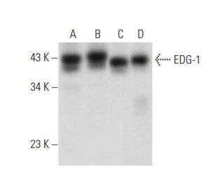
- EDG-1/S1P1/S1PR1 Antibody A-6 è un monoclonale di topo IgG3 κ EDG-1/S1P1/S1PR1 antibody, citato in 23 pubblicazioni, fornito in 200 µg/ml
- generato contro gli amminoacidi 322-381 di EDG-1 di origine human
- raccomandato per il rilevamento di EDG-1 di origine human in WB, IP, IF, IHC(P) e ELISA
- disponibile coniugato ad agarosio for IP; HRP for WB, IHC(P) and ELISA; phycoerythrin, fluorescein, Alexa Fluor® 488 or Alexa Fluor® 647 per IF, IHC(P) e FCM
- Al momento non abbiamo ancora completato l'identificazione dei reagenti secondari di rilevazione preferiti per EDG-1/S1P1/S1PR1 l'anticorpo (A-6). Questo lavoro è in corso.

LINK RAPIDI
VEDI ANCHE...
L'anticorpo EDG-1 (A-6) è un anticorpo monoclonale di topo IgG3 a catena leggera kappa che rileva EDG-1 di origine umana mediante western blotting (WB), immunoprecipitazione (IP), immunofluorescenza (IF), immunoistochimica e saggio di immunoassorbimento enzimatico (ELISA). L'anticorpo EDG-1 (A-6) è disponibile sia in forma non coniugata che in varie forme coniugate, tra cui agarosio, perossidasi di rafano (HRP), ficoeritrina (PE), isotiocianato di fluoresceina (FITC) e molteplici coniugati Alexa Fluor®. EDG-1, noto anche come S1PR1 o S1P1, è un membro della famiglia del gene di differenziazione endoteliale (EDG) dei recettori accoppiati a proteine G, che svolgono un ruolo cruciale nel mediare gli effetti delle molecole di segnalazione dei lisofosfolipidi, come la sfingosina-1-fosfato (S1P) e l'acido lisofosfatidico (LPA). L'EDG-1 si trova principalmente sulla superficie cellulare ed è coinvolto in vari processi cellulari, tra cui la sopravvivenza, la crescita, la migrazione e la differenziazione delle cellule, accoppiandosi alle proteine Gi per attivare le vie di segnalazione a valle, come Akt. Ciò è particolarmente importante nelle cellule di glioma, dove l'espressione di EDG-1 è associata alla progressione e alla sopravvivenza del tumore. La capacità di EDG-1 di interagire con più proteine G influenza un'ampia gamma di risposte fisiologiche, rendendo EDG-1 un bersaglio significativo per la ricerca nella biologia del cancro e nello sviluppo terapeutico.
Alexa Fluor® è un marchio registrato di Molecular Probes Inc., OR., USA
LI-COR® e Odyssey® sono marchi registrati di LI-COR Biosciences
Anticorpo EDG-1/S1P1/S1PR1 (A-6) Riferimenti:
- Proprietà farmacologiche e trasduzione del segnale differenziate dei recettori della sfingosina 1-fosfato EDG-1, EDG-3 e EDG-5. | Ancellin, N. and Hla, T. 1999. J Biol Chem. 274: 18997-9002. PMID: 10383399
- Una sottofamiglia di recettori cellulari accoppiati a proteine G per lisofosfolipidi e lisosfingolipidi. | Goetzl, EJ. and An, S. 1999. Adv Exp Med Biol. 469: 259-64. PMID: 10667339
- La sfingosina-1-fosfato è un ligando per il recettore accoppiato a proteine G EDG-6. | Van Brocklyn, JR., et al. 2000. Blood. 95: 2624-9. PMID: 10753843
- Segnalazione della sfingosina-1-fosfato attraverso la famiglia EDG-1 di recettori accoppiati a proteine G. | Hla, T., et al. 2000. Ann N Y Acad Sci. 905: 16-24. PMID: 10818438
- Sfingosina-1-fosfato: un ligando per la famiglia EDG-1 dei recettori accoppiati a proteine G. | Spiegel, S. 2000. Ann N Y Acad Sci. 905: 54-60. PMID: 10818441
- Segnalazione della sfingosina 1-fosfato nelle cellule di mammifero. | Pyne, S. and Pyne, NJ. 2000. Biochem J. 349: 385-402. PMID: 10880336
- Ruolo differenziale di Edg-1 e Edg-5, recettori della sfingosina 1-fosfato, nelle vie di segnalazione delle cellule di glioma C6. | Sato, K., et al. 2000. Brain Res Mol Brain Res. 85: 151-60. PMID: 11146117
- Effetti selettivi del recettore dell'acido lisofosfatidico sulla migrazione delle cellule T Jurkat attraverso una membrana basale modello Matrigel. | Zheng, Y., et al. 2001. J Immunol. 166: 2317-22. PMID: 11160288
- Distribuzione del recettore Edg2 nel cervello di ratto adulto. | Handford, EJ., et al. 2001. Neuroreport. 12: 757-60. PMID: 11277579
- La sfingosina 1-fosfato attiva Akt, la produzione di ossido nitrico e la chemiotassi attraverso un percorso proteina Gi/fosfoinositide 3-chinasi nelle cellule endoteliali. | Morales-Ruiz, M., et al. 2001. J Biol Chem. 276: 19672-7. PMID: 11278592
- Ruolo della N-glicosilazione nella dinamica di Edg-1/S1P1 in cellule stimolate dalla sfingosina 1-fosfato. | Kohno, T. and Igarashi, Y. 2004. Glycoconj J. 21: 497-501. PMID: 15750791
Informazioni ordini
| Nome del prodotto | Codice del prodotto | UNITÀ | Prezzo | Quantità | Preferiti | |
EDG-1/S1P1/S1PR1 Anticorpo (A-6) | sc-48356 | 200 µg/ml | $322.00 | |||
EDG-1/S1P1/S1PR1 Anticorpo (A-6) AC | sc-48356 AC | 500 µg/ml, 25% agarose | $424.00 | |||
EDG-1/S1P1/S1PR1 Anticorpo (A-6) Alexa Fluor® 488 | sc-48356 AF488 | 200 µg/ml | $364.00 | |||
EDG-1/S1P1/S1PR1 Anticorpo (A-6) Alexa Fluor® 647 | sc-48356 AF647 | 200 µg/ml | $364.00 | |||
EDG-1/S1P1/S1PR1 Anticorpo (A-6) FITC | sc-48356 FITC | 200 µg/ml | $336.00 | |||
EDG-1/S1P1/S1PR1 Anticorpo (A-6) HRP | sc-48356 HRP | 200 µg/ml | $322.00 | |||
EDG-1/S1P1/S1PR1 Anticorpo (A-6) PE | sc-48356 PE | 200 µg/ml | $349.00 |