")
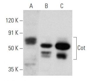
: sc-373677. Western blot analysis of Cot expression in Jurkat (A), NIH/3T3 (B) and RAW 264.7 (C) whole cell lysates.")
: sc-373677. Western blot analysis of Cot expression in AML-193 whole cell lysate.")
: sc-373677. Western blot analysis of Cot expression in CTLL-2 whole cell lysate.")
Cot Antibody (H-7): sc-373677
- Cot Antibody (H-7) is a mouse monoclonal IgG1 κ Cot antibody, cited in 9 publications, provided at 200 µg/ml
- specific for an epitope mapping between amino acids 441-467 at the C-terminus of Cot of mouse origin
- Cot Antibody (H-7) is recommended for detection of Cot of mouse, rat and human origin by WB, IP, IF and ELISA
- Anti-Cot Antibody (H-7) is available conjugated to agarose for IP; HRP for WB, IHC(P) and ELISA; and to either phycoerythrin or FITC for IF, IHC(P) and FCM
- also available conjugated to Alexa Fluor® 488, Alexa Fluor® 546, Alexa Fluor® 594 or Alexa Fluor® 647 for WB (RGB), IF, IHC(P) and FCM, and for use with RGB fluorescent imaging systems, such as iBright™ FL1000, FluorChem™, Typhoon, Azure and other comparable systems
- also available conjugated to Alexa Fluor® 680 or Alexa Fluor® 790 for WB (NIR), IF and FCM; for use with Near-Infrared (NIR) detection systems, such as LI-COR®Odyssey®, iBright™ FL1000, FluorChem™, Typhoon, Azure and other comparable systems
- m-IgG Fc BP-HRP, m-IgG1 BP-HRP and m-IgGκ BP-HRP are the preferred secondary detection reagents for Cot Antibody (H-7) for WB applications. These reagents are now offered in bundles with Cot Antibody (H-7) (see ordering information below).

QUICK LINKS
SEE ALSO...
Cot Antibody (H-7) is a mouse monoclonal IgG1 kappa light chain antibody that detects Cot protein of mouse, rat, and human origin by western blotting (WB), immunoprecipitation (IP), immunofluorescence (IF), and enzyme-linked immunosorbent assay (ELISA). anti-Cot antibody (H-7) is available in both non-conjugated and various conjugated forms, including agarose, horseradish peroxidase (HRP), phycoerythrin (PE), fluorescein isothiocyanate (FITC), and multiple Alexa Fluor® conjugates. Cot protein, also known as mitogen-activated protein kinase kinase kinase 8, plays a crucial role in cell signaling pathways, particularly in activating the mitogen-activated protein kinase (MAPK) pathway, which regulates cellular processes like proliferation, differentiation, and survival. Cot protein is primarily located in the cytoplasm, where Cot interacts with other signaling molecules to modulate cellular responses to external stimuli. Notably, Cot has been implicated in T lymphocyte activation, highlighting Cot′s importance in immune responses. Additionally, Cot protein undergoes post-translational modifications that influence activity and interactions, further underscoring Cot′s role in cellular signaling networks. Overexpression of Cot induces MAP kinase activation, and Cot′s function is intricately linked to proteins such as Ras and Raf-1, suggesting Cot may form a multimeric complex that regulates downstream signaling events. This complex interplay of interactions and modifications makes Cot a significant target for research in cancer biology and immunology.
Alexa Fluor® is a trademark of Molecular Probes Inc., OR., USA
LI-COR® and Odyssey® are registered trademarks of LI-COR Biosciences
Cot Antibody (H-7) References:
- Identification of a novel human kinase supporter of Ras (hKSR-2) that functions as a negative regulator of Cot (Tpl2) signaling. | Channavajhala, PL., et al. 2003. J Biol Chem. 278: 47089-97. PMID: 12975377
- Cell biology. A signal chain of events. | Roberts, TM. 1992. Nature. 360: 534-5. PMID: 1334231
- Ras p21: effects and regulation. | Haubruck, H. and McCormick, F. 1991. Biochim Biophys Acta. 1072: 215-29. PMID: 1751548
- Cot/Tpl-2 protein kinase as a target for the treatment of inflammatory disease. | George, D. and Salmeron, A. 2009. Curr Top Med Chem. 9: 611-22. PMID: 19689369
- COT drives resistance to RAF inhibition through MAP kinase pathway reactivation. | Johannessen, CM., et al. 2010. Nature. 468: 968-72. PMID: 21107320
- Cot/tpl2 participates in the activation of macrophages by adiponectin. | Sanz-Garcia, C., et al. 2014. J Leukoc Biol. 95: 917-30. PMID: 24532642
- Tumor progression locus 2 (TPL2): A Cot-plicated progression from inflammation to chronic liver disease. | Gutierrez, AH., et al. 2023. Biochim Biophys Acta Mol Basis Dis. 1869: 166660. PMID: 36764206
- Tpl-2 acts in concert with Ras and Raf-1 to activate mitogen-activated protein kinase. | Patriotis, C., et al. 1994. Proc Natl Acad Sci U S A. 91: 9755-9. PMID: 7937886
- The human cot proto-oncogene encodes two protein serine/threonine kinases with different transforming activities by alternative initiation of translation. | Aoki, M., et al. 1993. J Biol Chem. 268: 22723-32. PMID: 8226782
- Requirement for Raf and MAP kinase function during the meiotic maturation of Xenopus oocytes. | Fabian, JR., et al. 1993. J Cell Biol. 122: 645-52. PMID: 8335690
- The MAP kinase cascade is essential for diverse signal transduction pathways. | Nishida, E. and Gotoh, Y. 1993. Trends Biochem Sci. 18: 128-31. PMID: 8388132
- Cot kinase activates tumor necrosis factor-alpha gene expression in a cyclosporin A-resistant manner. | Ballester, A., et al. 1998. J Biol Chem. 273: 14099-106. PMID: 9603908
Ordering Information
| Product Name | Catalog # | UNIT | Price | Qty | FAVORITES | |
Cot Antibody (H-7) | sc-373677 | 200 µg/ml | $322.00 | |||
Cot Antibody (H-7): m-IgG Fc BP-HRP Bundle | sc-529536 | 200 µg Ab; 10 µg BP | $361.00 | |||
Cot Antibody (H-7): m-IgGκ BP-HRP Bundle | sc-522456 | 200 µg Ab, 40 µg BP | $361.00 | |||
Cot Antibody (H-7): m-IgG1 BP-HRP Bundle | sc-543564 | 200 µg Ab; 20 µg BP | $361.00 | |||
Cot Antibody (H-7) AC | sc-373677 AC | 500 µg/ml, 25% agarose | $424.00 | |||
Cot Antibody (H-7) HRP | sc-373677 HRP | 200 µg/ml | $322.00 | |||
Cot Antibody (H-7) FITC | sc-373677 FITC | 200 µg/ml | $336.00 | |||
Cot Antibody (H-7) PE | sc-373677 PE | 200 µg/ml | $349.00 | |||
Cot Antibody (H-7) Alexa Fluor® 488 | sc-373677 AF488 | 200 µg/ml | $364.00 | |||
Cot Antibody (H-7) Alexa Fluor® 546 | sc-373677 AF546 | 200 µg/ml | $364.00 | |||
Cot Antibody (H-7) Alexa Fluor® 594 | sc-373677 AF594 | 200 µg/ml | $364.00 | |||
Cot Antibody (H-7) Alexa Fluor® 647 | sc-373677 AF647 | 200 µg/ml | $364.00 | |||
Cot Antibody (H-7) Alexa Fluor® 680 | sc-373677 AF680 | 200 µg/ml | $364.00 | |||
Cot Antibody (H-7) Alexa Fluor® 790 | sc-373677 AF790 | 200 µg/ml | $364.00 | |||
Cot (H-7) Neutralizing Peptide | sc-373677 P | 100 µg/0.5 ml | $69.00 |