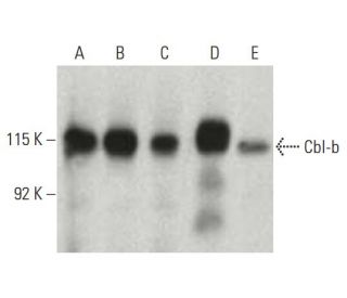
Date published: 2025-9-7

00800 4573 8000

SCBT Portrait Logo
Seach Input

Cbl-b Antikörper (G-1): sc-8006

4.4(17)
Produkt bewertenBitte stellen Sie eine Frage

Datenblätter
  • Cbl-b Antikörper G-1 ist ein Maus monoklonales IgG1 κ Cbl-b Antikörper, verwendet in 86 wissenschaftlichen Veröffentlichungen, in einer Menge von 200 µg/ml
  • gezogen gegen Aminosäuren 29-483 gelegen am N-terminus von Cbl-b aus der Spezies human
  • Cbl-b Antikörper (G-1) ist empfohlen für die Detektion von Cbl-b aus der Spezies mouse, rat und human per WB, IP, IF, IHC(P) und ELISA
  • Anti-Cbl-b Antikörper (G-1) ist erhältlich als Konjugat mit Agarose für IP; HRP für WB, IHC(P) und ELISA; und entweder mit Phycoerythrin oder FITC für IF, IHC(P) und FCM
  • auch erhältlich als Konjugat mit Alexa Fluor® 488, Alexa Fluor® 546, Alexa Fluor® 594 oder Alexa Fluor® 647 für IF, IHC(P) und FCM
  • auch erhältlich als Konjugat mit Alexa Fluor® 680 oder Alexa Fluor® 790 für WB (NIR), IF und FCM
  • Bitte kontaktieren Sie uns um ein 10 µg GRATIS Sample vom Cbl-b (G-1): sc-8006 zu erhalten.
  • m-IgG Fc BP-HRP, m-IgG1 BP-HRP und m-IgGκ BP-HRP sind die empfohlenen Detektionsreagenzien für Cbl-b Antikörper (G-1) für WB and IHC(P) Experimente. Diese Reagenzien werden nun auch als Bundle angeboten, zusammen mit Cbl-b Antikörper (G-1) (Bestellinformation unten stehend).

Direktverknüpfungen

Siehe auch...

Der Cbl-b-Antikörper (G-1) ist ein monoklonaler IgG1-Kappa-Leichtketten-Antikörper der Maus, der das Cbl-b-Protein von Mäusen, Ratten und Menschen durch Western Blot (WB), Immunopräzipitation (IP), Immunfluoreszenz (IF), Immunhistochemie und Enzyme-linked Immunosorbent Assay (ELISA) nachweist. Der monoklonale Cbl-b-Antikörper (G-1) ist sowohl in nicht konjugierter als auch in verschiedenen konjugierten Formen erhältlich, darunter Agarose, Meerrettichperoxidase (HRP), Phycoerythrin (PE), Fluoresceinisothiocyanat (FITC) und mehrere Alexa Fluor®-Konjugate. Das Cbl-b-Protein, auch bekannt als RING-Finger-Protein 56, spielt eine entscheidende Rolle bei der Regulierung von Signaltransduktionswegen, indem es als E3-Ubiquitin-Ligase fungiert, die für die Ubiquitinierung und den anschließenden Abbau von Zielproteinen unerlässlich ist. Diese Regulierung ist besonders wichtig für die Aufrechterhaltung der zellulären Homöostase und die Verhinderung einer unkontrollierten Zellproliferation, die zu Krebs führen kann. Cbl-b wird in einer Vielzahl von Geweben exprimiert, darunter in normalen und bösartigen Brustepithelzellen sowie in hämatopoetischen Geweben, was seine Bedeutung sowohl für die normale Physiologie als auch für Krankheitszustände unterstreicht. Das Protein enthält eine prolinreiche Domäne, ein Kernlokalisierungssignal, einen C3HC4-Zinkfinger und einen mutmaßlichen Leucin-Zipper, die für seine Wechselwirkungen mit anderen Signalproteinen entscheidend sind. Bemerkenswert ist, dass sich das Cbl-b-Gen in einer Region des menschlichen Chromosoms 11 befindet, die häufig an Translokationen und Deletionen im Zusammenhang mit verschiedenen Leukämien beteiligt ist, was die Bedeutung von Cbl-b in der Krebsbiologie unterstreicht.

Ausschließlich für Forschungszwecke. Nicht für diagnostische oder therapeutische Zwecke bestimmt.

Alexa Fluor® ist ein Markenzeichen von Molecular Probes Inc., OR., USA

LI-COR® und Odyssey® sind Markenzeichen von LI-COR Biosciences

Cbl-b Antikörper (G-1) Literaturhinweise:

  1. Die Sequenzen der c-cbl-Proto-Onkogene von Mensch und Maus zeigen, dass v-cbl durch eine große Trunkierung erzeugt wurde, die eine prolinreiche Domäne und ein Leucin-Zipper-ähnliches Motiv umfasst.  |  Blake, TJ., et al. 1991. Oncogene. 6: 653-7. PMID: 2030914
  2. Das Proto-Onkogen c-cbl wird bevorzugt in Thymus- und Hodengewebe exprimiert und kodiert für ein Kernprotein.  |  Langdon, WY., et al. 1989. J Virol. 63: 5420-4. PMID: 2585608
  3. Identifizierung von zwei murinen Loci, die dem v-cbl-Onkogen homolog sind.  |  Regnier, DC., et al. 1989. J Virol. 63: 3678-82. PMID: 2760978
  4. v-cbl, ein Onkogen aus einem dual-rekombinanten murinen Retrovirus, das frühe B-Linien-Lymphome hervorruft.  |  Langdon, WY., et al. 1989. Proc Natl Acad Sci U S A. 86: 1168-72. PMID: 2784003
  5. Die sequenzielle Ubiquitinierung von NLRP3 durch RNF125 und Cbl-b begrenzt die Aktivierung des Inflammasoms und die Endotoxämie.  |  Tang, J., et al. 2020. J Exp Med. 217: PMID: 31999304
  6. Ubiquitin-Ligase Cbl-b und inhibitorische Cblin-Peptide.  |  Nikawa, T. and Ishidoh, K. 2020. Biochim Biophys Acta Proteins Proteom. 1868: 140495. PMID: 32663526
  7. Die Deletion von Cbl-b hemmt die Erschöpfung der CD8+-T-Zellen und fördert die CAR-T-Zell-Funktion.  |  Kumar, J., et al. 2021. J Immunother Cancer. 9: PMID: 33462140
  8. Cbl-b als Ziel bei der Krebsimmuntherapie.  |  Augustin, RC., et al. 2023. J Immunother Cancer. 11: PMID: 36750253
  9. Cbl und Cbl-b regulieren EGFR unabhängig voneinander durch unterschiedliche Rezeptorinteraktionsmodi.  |  Pinilla-Macua, I. and Sorkin, A. 2023. Mol Biol Cell. 34: ar134. PMID: 37903221
  10. CBL-B – ein aufstrebendes immunonkologisches Ziel.  |  Fusco, R., et al. 2025. Expert Opin Ther Pat. 35: 47-64. PMID: 39582379
  11. Klonierung und Charakterisierung von cbl-b: ein SH3-bindendes Protein mit Homologie zum c-cbl-Proto-Onkogen.  |  Keane, MM., et al. 1995. Oncogene. 10: 2367-77. PMID: 7784085
  12. Bildung von c-Cbl.Phosphatidylinositol-3-Kinase-Komplexen auf Lymphozytenmembranen durch einen p56lck-unabhängigen Mechanismus.  |  Hartley, D. and Corvera, S. 1996. J Biol Chem. 271: 21939-43. PMID: 8702998

Bestellinformation

ProduktKatalog #EINHEITPreisANZAHLFavoriten

Cbl-b Antikörper (G-1)

sc-8006
200 µg/ml
$316.00

Cbl-b (G-1): m-IgG Fc BP-HRP Bundle

sc-528197
200 µg Ab; 10 µg BP
$354.00

Cbl-b (G-1): m-IgGκ BP-HRP Bundle

sc-520519
200 µg Ab, 40 µg BP
$354.00

Cbl-b (G-1): m-IgG1 BP-HRP Bundle

sc-542810
200 µg Ab; 20 µg BP
$354.00

Cbl-b Antikörper (G-1) AC

sc-8006 AC
500 µg/ml, 25% agarose
$416.00

Cbl-b Antikörper (G-1) HRP

sc-8006 HRP
200 µg/ml
$316.00

Cbl-b Antikörper (G-1) FITC

sc-8006 FITC
200 µg/ml
$330.00

Cbl-b Antikörper (G-1) PE

sc-8006 PE
200 µg/ml
$343.00

Cbl-b Antikörper (G-1) Alexa Fluor® 488

sc-8006 AF488
200 µg/ml
$357.00

Cbl-b Antikörper (G-1) Alexa Fluor® 546

sc-8006 AF546
200 µg/ml
$357.00

Cbl-b Antikörper (G-1) Alexa Fluor® 594

sc-8006 AF594
200 µg/ml
$357.00

Cbl-b Antikörper (G-1) Alexa Fluor® 647

sc-8006 AF647
200 µg/ml
$357.00

Cbl-b Antikörper (G-1) Alexa Fluor® 680

sc-8006 AF680
200 µg/ml
$357.00

Cbl-b Antikörper (G-1) Alexa Fluor® 790

sc-8006 AF790
200 µg/ml
$357.00

Since it's marked for use for IP, I tried doing the IP using this G1 antibody followed by Western Blot for pTyrosine (PY20) and it didn't work whereas it has worked in reverse IP using PY20 followed by western blot for Cbl-b. Any advice? 

Gefragt von: LeilaniC
Thank you for your question. Cbl-b Antibody (G-1): sc-8006 is recommended for immunoprecipitation of Cbl-b. It would depend on your samples whether or not you could detect phosphorylated tyrosine by Western blot, following immunoprecipitation of Cbl.b. We would suggest doing a Western blot for Cbl-b in your samples before, and after IP with the (G-1) clone to determine whether or not the antibody is working for enrichment of Cbl-b. If you still have questions, please contact our Technical Service Department and we would be happy to help you troubleshoot these antibodies. Please call us toll-free at (800) 457-3801 option 2, or e-mail scbt@scbt.com.
Beantwortet von: Technical Support
Veröffentlichungsdatum: 2017-04-06

Hello, i have been working with C-20 antibody against Cbl-b in Jurkat cells for IP in the last years, and it worked very good (an excellent ab). But now, i have tested G-1 antibody and it didn't work, even with Cblb-overexpressing Jurkat. Any advice?

Gefragt von: Javier Robles
Thank you for your question. We are sorry to hear that this antibody did not perform for you as expected. We want you to be successful in your research. Santa Cruz Biotechnology does warrant our antibodies for one year from the date of purchase. Please contact our Technical Service Department (spain@scbt.com). We are happy to help you to troubleshoot the experiment.
Beantwortet von: Technical Support Europe
Veröffentlichungsdatum: 2017-01-31

What is the recommended secondary reagent to be used with this mouse monoclonal primary?

Gefragt von: DefinitelyNotMatt
Thank you for your question. We recommend using one our exclusive Mouse IgG Binding Proteins as a secondary detection reagent. A complete list of available binding proteins is available on our website here: https://www.scbt.com/scbt/browse/support-products-mouse-igg-binding-proteins/_/N-ecrety Please contact Technical Service if you have further questions concerning this product.
Beantwortet von: Tech Service 9
Veröffentlichungsdatum: 2017-01-04
  • y_2025, m_8, d_27, h_5CST
  • bvseo_bulk, prod_bvqa, vn_bulk_3.0.42
  • cp_1, bvpage1
  • co_hasquestionsanswers, tq_3
  • loc_de_DE, sid_8006, prod, sort_[SortEntry(order=LAST_APPROVED_ANSWER_SUBMISSION_TIME, direction=DESCENDING)]
  • clientName_scbt
  • bvseo_sdk, java_sdk, bvseo-4.0.0
  • CLOUD, getContent, 125ms
  • QUESTIONS, PRODUCT
Rated 5 von 5 von aus Nice productWorks well in primary mouse T cells and breast cancer cell lines in Western Blot and Immunoprecipitation. Consider using a low dilution to improve results (1:100)
Veröffentlichungsdatum: 2021-06-21
Rated 4 von 5 von aus worked well in western blotThis antibody worked well in western blot on mouse osteoclasts extract.
Veröffentlichungsdatum: 2017-10-25
Rated 5 von 5 von aus Good bandGood band seen in WB using human recombinant Cbl-b.
Veröffentlichungsdatum: 2017-05-07
Rated 5 von 5 von aus Great WB antibodyProduced specific results. Validated specificity with siRNA knockout. Highly recommended!
Veröffentlichungsdatum: 2017-01-10
Rated 5 von 5 von aus Great antibodyVery clear strong western data; low background;nice si rna control
Veröffentlichungsdatum: 2016-12-31
Rated 1 von 5 von aus No detectable expression in ARPE-19 cellsNo detectable expression in ARPE-19 cells after reaching confluence. Used 1:500 dilution, observed just non-specific bands.
Veröffentlichungsdatum: 2016-09-26
Rated 5 von 5 von aus Published in IF for paraffinPublished in IF for paraffin-embedded human T cells. -SCBT Publication Review
Veröffentlichungsdatum: 2015-05-11
Rated 4 von 5 von aus Great data published on Western blot on mouse 3T3Great data published on Western blot on mouse 3T3-L1 adipocytes. -SCBT Publication Review
Veröffentlichungsdatum: 2015-04-25
  • y_2025, m_8, d_27, h_5
  • bvseo_bulk, prod_bvrr, vn_bulk_3.0.42
  • cp_1, bvpage1
  • co_hasreviews, tv_0, tr_17
  • loc_de_DE, sid_8006, prod, sort_[SortEntry(order=SUBMISSION_TIME, direction=DESCENDING)]
  • clientName_scbt
  • bvseo_sdk, java_sdk, bvseo-4.0.0
  • CLOUD, getReviews, 40ms
  • REVIEWS, PRODUCT
Cbl-b Antikörper (G-1) wurde bewertet mit 4.4 von 5 von 17.
  • y_2025, m_8, d_27, h_5
  • bvseo_bulk, prod_bvrr, vn_bulk_3.0.42
  • cp_1, bvpage1
  • co_hasreviews, tv_0, tr_17
  • loc_de_DE, sid_8006, prod, sort_[SortEntry(order=SUBMISSION_TIME, direction=DESCENDING)]
  • clientName_scbt
  • bvseo_sdk, java_sdk, bvseo-4.0.0
  • CLOUD, getAggregateRating, 116ms
  • REVIEWS, PRODUCT