")
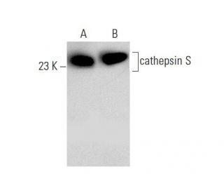
cathepsin S Antikörper (E-3): sc-271619
- cathepsin S Antikörper E-3 ist ein Maus monoklonales IgG1 κ cathepsin S Antikörper, verwendet in 35 wissenschaftlichen Veröffentlichungen, in einer Menge von 200 µg/ml
- spezifisch für ein Epitop, welches zwischen den Aminosäuren 302-331 am C-terminus von cathepsin S aus der Spezies human liegt
- cathepsin S Antikörper (E-3) ist empfohlen für die Detektion von cathepsin S aus der Spezies mouse, rat und human per WB, IP, IF, IHC(P) und ELISA
- Anti-cathepsin S Antikörper (E-3) ist erhältlich als Konjugat mit Agarose für IP; HRP für WB, IHC(P) und ELISA; und entweder mit Phycoerythrin oder FITC für IF, IHC(P) und FCM
- auch erhältlich als Konjugat mit Alexa Fluor® 488, Alexa Fluor® 546, Alexa Fluor® 594 oder Alexa Fluor® 647 für IF, IHC(P) und FCM
- auch erhältlich als Konjugat mit Alexa Fluor® 680 oder Alexa Fluor® 790 für WB (NIR), IF und FCM
- m-IgG Fc BP-HRP und m-IgG1 BP-HRP sind die bevorzugten sekundären Nachweisreagenzien für cathepsin S Antikörper (E-3) for WB and IHC(P) applications. Diese Reagenzien werden jetzt in Bündeln mit cathepsin S Antikörper (E-3) angeboten(siehe Bestellinformationen unten).

Direktverknüpfungen
Siehe auch...
Die Kathepsin-Familie von proteolytischen Enzymen enthält mehrere unterschiedliche Klassen von Proteasen. Die Cystein-Protease-Klasse umfasst Kathepsine B, L, H, K, S und O. Die Aspartyl-Protease-Klasse besteht aus Kathepsinen D und E. Kathepsin G ist in der Serin-Protease-Klasse. Die meisten Kathepsine sind lysosomal und jede ist an der Zellmetabolismus beteiligt und nimmt an verschiedenen Ereignissen wie Peptidbiosynthese und Proteinabbau teil. Cathepsin S wurde als eine elastinolytische Cystein-Protease in aveloaren Makrophagen gezeigt. Ein IgG1 κ Maus-Monoklonales Cathepsin S Antikörper (E-3) wird angeboten, das das Cathepsin S Protein von Maus-, Ratte- und menschlicher Herkunft durch WB, IP, IF, IHC (P) und ELISA detektiert. Cathepsin S Antikörper (E-3) ist sowohl in der nicht konjugierten Form als auch in mehreren konjugierten Formen des Cathepsin S Antikörpers erhältlich, einschließlich Agarose, HRP, PE, FITC und mehreren Alexa Fluor® Konjugaten.
Alexa Fluor® ist ein Markenzeichen von Molecular Probes Inc., OR., USA
LI-COR® und Odyssey® sind Markenzeichen von LI-COR Biosciences
cathepsin S Antikörper (E-3) Literaturhinweise:
- Molekulare Klonierung und Expression von menschlichem alveolärem Makrophagen-Cathepsin S, einer elastinolytischen Cysteinprotease. | Shi, GP., et al. 1992. J Biol Chem. 267: 7258-62. PMID: 1373132
- Molekulare Organisation des menschlichen Cathepsin-D-Gens. | Redecker, B., et al. 1991. DNA Cell Biol. 10: 423-31. PMID: 2069717
- Cathepsin S als Ziel bei Magenkrebs. | da Costa, AC., et al. 2020. Mol Clin Oncol. 12: 99-103. PMID: 31929878
- Elafin kehrt die intestinale Fibrose durch Hemmung des Cathepsin S-vermittelten Protease-aktivierten Rezeptors 2 um. | Xie, Y., et al. 2022. Cell Mol Gastroenterol Hepatol. 14: 841-876. PMID: 35840034
- Die Hemmung von Cathepsin S unterdrückt die experimentelle systemische Lupus erythematodes-assoziierte pulmonale arterielle Remodellierung. | Yen, TH., et al. 2022. Int J Mol Sci. 23: PMID: 36293172
- Molekulare Klonierung und Sequenzierung der cDNA für Ratten-Cathepsin L. | Ishidoh, K., et al. 1987. FEBS Lett. 223: 69-73. PMID: 3666143
- Molekulare Klonierung und Sequenzierung der cDNA für Ratten-Cathepsin H. Homologie in Pro-Peptid-Regionen von Cysteinproteinasen. | Ishidoh, K., et al. 1987. FEBS Lett. 226: 33-7. PMID: 3691815
- Isolierung und Sequenzierung von zwei cDNA-Klonen, die für Rattenmilz-Cathepsin E kodieren, und Analyse der Aktivierung von gereinigtem Procathepsin E. | Okamoto, K., et al. 1995. Arch Biochem Biophys. 322: 103-11. PMID: 7574663
- Molekulare Klonierung von menschlichem Cathepsin O, einer neuen Endoproteinase und Homolog von Kaninchen OC2. | Shi, GP., et al. 1995. FEBS Lett. 357: 129-34. PMID: 7805878
- Cathepsin B, eine Cysteinprotease, die bei der Metastasenbildung eine Rolle spielt, wird auch bei der Rückbildung der Rattenprostata und der Brustdrüsen exprimiert. | Guenette, RS., et al. 1994. Eur J Biochem. 226: 311-21. PMID: 8001549
- Molekulare Klonierung, chromosomale Lage und gewebespezifische Expression des murinen Cathepsin-G-Gens. | Heusel, JW., et al. 1993. Blood. 81: 1614-23. PMID: 8453108
- Maus-Cathepsin K: cDNA-Klonierung und vorherrschende Expression des Gens in Osteoklasten und in einigen hypertrophierenden Chondrozyten während der Mausentwicklung. | Rantakokko, J., et al. 1996. FEBS Lett. 393: 307-13. PMID: 8814310
Bestellinformation
| Produkt | Katalog # | EINHEIT | Preis | ANZAHL | Favoriten | |
cathepsin S Antikörper (E-3) | sc-271619 | 200 µg/ml | $322.00 | |||
cathepsin S (E-3): m-IgG Fc BP-HRP Bundle | sc-527268 | 200 µg Ab; 10 µg BP | $361.00 | |||
cathepsin S (E-3): m-IgG1 BP-HRP Bundle | sc-532641 | 200 µg Ab; 20 µg BP | $361.00 | |||
cathepsin S Antikörper (E-3) AC | sc-271619 AC | 500 µg/ml, 25% agarose | $424.00 | |||
cathepsin S Antikörper (E-3) HRP | sc-271619 HRP | 200 µg/ml | $322.00 | |||
cathepsin S Antikörper (E-3) FITC | sc-271619 FITC | 200 µg/ml | $336.00 | |||
cathepsin S Antikörper (E-3) PE | sc-271619 PE | 200 µg/ml | $349.00 | |||
cathepsin S Antikörper (E-3) Alexa Fluor® 488 | sc-271619 AF488 | 200 µg/ml | $364.00 | |||
cathepsin S Antikörper (E-3) Alexa Fluor® 546 | sc-271619 AF546 | 200 µg/ml | $364.00 | |||
cathepsin S Antikörper (E-3) Alexa Fluor® 594 | sc-271619 AF594 | 200 µg/ml | $364.00 | |||
cathepsin S Antikörper (E-3) Alexa Fluor® 647 | sc-271619 AF647 | 200 µg/ml | $364.00 | |||
cathepsin S Antikörper (E-3) Alexa Fluor® 680 | sc-271619 AF680 | 200 µg/ml | $364.00 | |||
cathepsin S Antikörper (E-3) Alexa Fluor® 790 | sc-271619 AF790 | 200 µg/ml | $364.00 | |||
cathepsin S (E-3) Neutralizing Peptid | sc-271619 P | 100 µg/0.5 ml | $69.00 |