Date published: 2026-5-5

1-800-457-3801

SCBT Portrait Logo
Seach Input

p-Cofilin 1 Antibody (E-5): sc-271921

5.0(11)
Write a reviewAsk a question

Datasheets
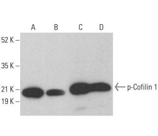
  • p-Cofilin 1 Antibody (E-5) is a mouse monoclonal IgM κ, cited in 13 publications, provided at 200 µg/ml
  • raised against a short amino acid sequence containing phosphorylated Ser 3 of Cofilin 1 of human origin
  • recommended for detection of Ser 3 phosphorylated Cofilin 1 of mouse, rat and human origin by WB, IP, IF, IHC(P) and ELISA; also reactive with additional species, including and bovine and porcine
  • m-IgGκ BP-HRP is the preferred secondary detection reagent for p-Cofilin 1 Antibody (E-5) for WB and IHC(P) applications. This reagent is now offered in a bundle with p-Cofilin 1 Antibody (E-5) (see ordering information below). For additional m-IgGκ BP conjugates see our complete list of Mouse IgG Binding Proteins.
Gene Editing Promo Banner

QUICK LINKS

SEE ALSO...

p-Cofilin 1 Antibody (E-5) is a mouse monoclonal IgM antibody that detects p-Cofilin 1, specifically the Ser 3 phosphorylated form, in mouse, rat, and human samples through applications such as western blotting (WB), immunoprecipitation (IP), immunofluorescence (IF), immunohistochemistry, and enzyme-linked immunosorbent assay (ELISA). p-Cofilin 1 Antibody (E-5) is available in a non-conjugated format, allowing for versatile experimental use. Cofilin, a critical actin-binding protein, plays a vital role in regulating actin cytoskeleton, which is essential for various cellular processes, including motility, division, and shape maintenance. Phosphorylation of cofilin at Serine 3 by LIM-kinase 1 (LIMK-1) inhibits actin-depolymerizing activity, thereby stabilizing actin filaments and promoting cytoskeletal integrity. This regulation allows cells to respond dynamically to signaling cues, facilitating processes such as cell migration and tissue remodeling. Cofilin′s interaction with other actin-binding proteins further underscores cofilin′s importance in maintaining the delicate balance of actin dynamics within cells, making anti-p-Cofilin 1 antibody (E-5) an invaluable tool for researchers studying cytoskeletal organization and its implications in health and disease.

For Research Use Only. Not Intended for Diagnostic or Therapeutic Use.

Ordering Information

Product NameCatalog #UNITPriceQtyFAVORITES

p-Cofilin 1 Antibody (E-5)

sc-271921
200 µg/ml
$322.00

p-Cofilin 1 Antibody (E-5): m-IgGκ BP-HRP Bundle

sc-551996
200 µg Ab; 40 µg BP
$361.00

p-Cofilin 1 (E-5) Neutralizing Peptide

sc-271921 P
100 µg/0.5 ml
$69.00

Has this antibody been tested on frozen tissue sections for immunofluorescence, specifically mouse tissues? Are any antigen retrieval steps recommended?

Asked by: HB23
Thank you for your question. sc-271921 is recommended for mouse, rat, human , bovine , and porcine. We validate this antibody for WB, IP, IF, IHC(P) and ELISA. Please see attached protocol for antigen retrieval. https://www.scbt.com/resources/protocols/immunofluorescence-cell-staining
Answered by: BlakeJ
Date published: 2023-03-16

Does this antibody (or any of the other anti-cofilin antibodies currently available) recognize Drosophila Cofilin by IF?

Asked by: JSR1
Thank you for your question. Sequence analysis shows very low to no homology between the epitope sequence of the antibodies and the target in Drosophila. If you have additional questions, please contact Technical Support at 1800-457-3801 option 2, by emailing scbt@scbt.com or by live chat on our website at www.scbt.com so we may further assist you.
Answered by: Santa Cruz TS
Date published: 2021-07-07

What application is the blocking peptide sc-271921 P appropriate for?

Asked by: Germaine
Thank you for your question. The blocking peptide is intended for use as a negative control, by pre-adsorbing the mouse monoclonal antibody against the antigen. For full protocol details, please contact our Technical Services Department or view our online protocol here: https://www.scbt.com/scbt/resources/protocols/peptide-neutralization
Answered by: Technical Support
Date published: 2017-02-24
  • y_2026, m_5, d_5, h_11CST
  • bvseo_bulk, prod_bvqa, vn_bulk_3.0.43
  • cp_1, bvpage1
  • co_hasquestionsanswers, tq_3
  • loc_en_US, sid_271921, prod, sort_[SortEntry(order=LAST_APPROVED_ANSWER_SUBMISSION_TIME, direction=DESCENDING)]
  • clientName_scbt
  • bvseo_sdk, java_sdk, bvseo-4.0.0
  • CLOUD, getContent, 104ms
  • QUESTIONS, PRODUCT
Rated 5 out of 5 by from Anti-p-Cofilin per analisi in western blotL'Ab è stato testato utilizzando lisati cellulari MCF-7 e MDA-MD-231, ottenuti in RIPA buffer. 15 ug di proteine sono state separate tramite SDS-Page e trasferite su un filtro di nitrocellulosa. Il bloccaggio è stato effettuato in Blotto A per 1h, l'Ab primario è stato utilizzato alla diluizione di 1:1000 in 10 mL di Blotto A per 1h. Il secondario 1:2000 in Blotto A per 1h. Il segnale ottenuto è pulito, con un'unica banda del perso molecolare atteso. La specificità del segnale è stata verificata utilizzando l'inibitore della chinasi ROCK, Y-27632.
Date published: 2024-07-23
Rated 5 out of 5 by from Great antibody for western blottingWe used 1:1000 on western blotting from NPCs, it give a nice and clear band.
Date published: 2018-07-19
Rated 5 out of 5 by from Great AbUsed a 1:300 dilution for WB with 1- SUM159, 2- Caco-2, and 3- 293-T cell lysates. detected a very clean and strong band.
Date published: 2018-01-30
Rated 5 out of 5 by from Works very well on WBI used this antibody on brain tissue (protein load: 30 uL, Ab concentration: 1/1000), and it worked very well: one band and good intensity for quantification.
Date published: 2018-01-08
Rated 5 out of 5 by from The band is present and abundantThis antibody worked great with HEK293 whole cell lysis at 1:500 dilution and incubation with 5% BSA
Date published: 2017-11-09
Rated 5 out of 5 by from good antibodyI used it at 1:150 dilution with milk and worked very well.
Date published: 2017-04-14
Rated 5 out of 5 by from Sensitive and SpecificGreat specificity to phosphorylated Cofilin 1 as shown by phosphatase treatment.
Date published: 2017-01-16
Rated 5 out of 5 by from Very potent and highly specific antibodyThis monoclonal antibody was independently tested by Kinexus Bioinformatics Corporation and determined to be an excellent antibody for Western blotting. Strong detection of cofilin 1 was observed, but no cross-reactivities detected by Western blotting in HeLa cells exposed to anisomycin for 2 hours. No detection of the cofilin 1 target was observed in overnight-serum starved human A431 cells that were treated for 5 minutes with epidermal growth factor. However, there was no significant cross-reactivities on these A431 cell immunoblots with any off-target proteins.
Date published: 2016-12-08
  • y_2026, m_5, d_5, h_11
  • bvseo_bulk, prod_bvrr, vn_bulk_3.0.43
  • cp_1, bvpage1
  • co_hasreviews, tv_0, tr_11
  • loc_en_US, sid_271921, prod, sort_[SortEntry(order=SUBMISSION_TIME, direction=DESCENDING)]
  • clientName_scbt
  • bvseo_sdk, java_sdk, bvseo-4.0.0
  • CLOUD, getReviews, 8ms
  • REVIEWS, PRODUCT
p-Cofilin 1 Antibody (E-5) is rated 5.0 out of 5 by 11.
  • y_2026, m_5, d_5, h_11
  • bvseo_bulk, prod_bvrr, vn_bulk_3.0.43
  • cp_1, bvpage1
  • co_hasreviews, tv_0, tr_11
  • loc_en_US, sid_271921, prod, sort_[SortEntry(order=SUBMISSION_TIME, direction=DESCENDING)]
  • clientName_scbt
  • bvseo_sdk, java_sdk, bvseo-4.0.0
  • CLOUD, getAggregateRating, 103ms
  • REVIEWS, PRODUCT