")
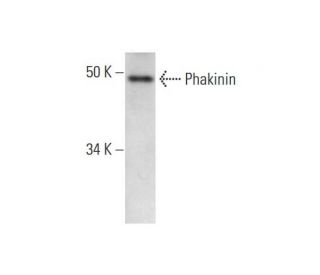
: sc-390848. Western blot analysis of Phakinin expression in mouse eye tissue extract.")
Phakinin Antibody (D-7): sc-390848
- Phakinin Antibody (D-7) is a mouse monoclonal IgG1 κ Phakinin antibody provided at 200 µg/ml
- specific for an epitope mapping between amino acids 319-340 within an internal region of Phakinin of mouse origin
- Phakinin Antibody (D-7) is recommended for detection of Phakinin of mouse and rat origin by WB, IP, IF and ELISA
- Anti-Phakinin Antibody (D-7) is available conjugated to agarose for IP; HRP for WB, IHC(P) and ELISA; and to either phycoerythrin or FITC for IF, IHC(P) and FCM
- also available conjugated to Alexa Fluor® 488, Alexa Fluor® 546, Alexa Fluor® 594 or Alexa Fluor® 647 for WB (RGB), IF, IHC(P) and FCM, and for use with RGB fluorescent imaging systems, such as iBright™ FL1000, FluorChem™, Typhoon, Azure and other comparable systems
- also available conjugated to Alexa Fluor® 680 or Alexa Fluor® 790 for WB (NIR), IF and FCM; for use with Near-Infrared (NIR) detection systems, such as LI-COR®Odyssey®, iBright™ FL1000, FluorChem™, Typhoon, Azure and other comparable systems
- m-IgG Fc BP-HRP is the preferred secondary detection reagent for Phakinin Antibody (D-7) for WB applications. This reagent is now offered in a bundle with Phakinin Antibody (D-7) (see ordering information below).

QUICK LINKS
SEE ALSO...
Phakinin Antibody (D-7) is a mouse monoclonal IgG1 kappa light chain antibody that detects Phakinin protein of mouse and rat origin by western blotting (WB), immunoprecipitation (IP), immunofluorescence (IF), and enzyme-linked immunosorbent assay (ELISA). anti-Phakinin antibody (D-7) is available in both non-conjugated and various conjugated forms, including agarose, horseradish peroxidase (HRP), phycoerythrin (PE), fluorescein isothiocyanate (FITC), and multiple Alexa Fluor® conjugates. Phakinin, also known as BFSP2 (beaded filament structural protein 2), CP47, CP49 (lens fiber beaded filament protein CP49), or LIFL-L (lens intermediate filament-like light), is a membrane-associated and cytoskeletal intermediate filament protein that plays a crucial role in the eye lens. This protein is essential for the assembly of beaded filaments and the formation of cytoskeletal networks, which are vital for maintaining the optical properties and transparency of the lens. Phakinin′s unique structure, characterized by the absence of a C-terminal tail domain, distinguishes Phakinin from most intermediate filaments and contributes to the formation of the beaded filament structures specific to the lens. Phakinin interacts with Filensin, another intermediate filament protein, to create the 10-nanometer filamentous structures that are integral to lens integrity. Mutations in the gene encoding Phakinin can lead to lens cataracts, highlighting Phakinin′s importance in ocular health.
Alexa Fluor® is a trademark of Molecular Probes Inc., OR., USA
LI-COR® and Odyssey® are registered trademarks of LI-COR Biosciences
Phakinin Antibody (D-7) References:
- Autosomal-dominant congenital cataract associated with a deletion mutation in the human beaded filament protein gene BFSP2. | Jakobs, PM., et al. 2000. Am J Hum Genet. 66: 1432-6. PMID: 10739768
- Knockout of the intermediate filament protein CP49 destabilises the lens fibre cell cytoskeleton and decreases lens optical quality, but does not induce cataract. | Sandilands, A., et al. 2003. Exp Eye Res. 76: 385-91. PMID: 12573667
- Characterization of a mutation in the lens-specific CP49 in the 129 strain of mouse. | Alizadeh, A., et al. 2004. Invest Ophthalmol Vis Sci. 45: 884-91. PMID: 14985306
- Bfsp2 mutation found in mouse 129 strains causes the loss of CP49' and induces vimentin-dependent changes in the lens fibre cell cytoskeleton. | Sandilands, A., et al. 2004. Exp Eye Res. 78: 875-89. PMID: 15037121
- Eye lens proteomics: from global approach to detailed information about phakinin and gamma E and F crystallin genes. | Hoehenwarter, W., et al. 2005. Proteomics. 5: 245-57. PMID: 15744838
- The C terminus of lens aquaporin 0 interacts with the cytoskeletal proteins filensin and CP49. | Lindsey Rose, KM., et al. 2006. Invest Ophthalmol Vis Sci. 47: 1562-70. PMID: 16565393
- Insights into the beaded filament of the eye lens. | Perng, MD., et al. 2007. Exp Cell Res. 313: 2180-8. PMID: 17490642
- A role for lengsin, a recruited enzyme, in terminal differentiation in the vertebrate lens. | Wyatt, K., et al. 2008. J Biol Chem. 283: 6607-15. PMID: 18178558
Ordering Information
| Product Name | Catalog # | UNIT | Price | Qty | FAVORITES | |
Phakinin Antibody (D-7) | sc-390848 | 200 µg/ml | $322.00 | |||
Phakinin Antibody (D-7): m-IgG Fc BP-HRP Bundle | sc-526198 | 200 µg Ab; 10 µg BP | $361.00 | |||
Phakinin Antibody (D-7) AC | sc-390848 AC | 500 µg/ml, 25% agarose | $424.00 | |||
Phakinin Antibody (D-7) HRP | sc-390848 HRP | 200 µg/ml | $322.00 | |||
Phakinin Antibody (D-7) FITC | sc-390848 FITC | 200 µg/ml | $336.00 | |||
Phakinin Antibody (D-7) PE | sc-390848 PE | 200 µg/ml | $349.00 | |||
Phakinin Antibody (D-7) Alexa Fluor® 488 | sc-390848 AF488 | 200 µg/ml | $364.00 | |||
Phakinin Antibody (D-7) Alexa Fluor® 546 | sc-390848 AF546 | 200 µg/ml | $364.00 | |||
Phakinin Antibody (D-7) Alexa Fluor® 594 | sc-390848 AF594 | 200 µg/ml | $364.00 | |||
Phakinin Antibody (D-7) Alexa Fluor® 647 | sc-390848 AF647 | 200 µg/ml | $364.00 | |||
Phakinin Antibody (D-7) Alexa Fluor® 680 | sc-390848 AF680 | 200 µg/ml | $364.00 | |||
Phakinin Antibody (D-7) Alexa Fluor® 790 | sc-390848 AF790 | 200 µg/ml | $364.00 | |||
Phakinin (D-7) Neutralizing Peptide | sc-390848 P | 100 µg/0.5 ml | $69.00 |