")
Glycophorin A Antibody (R10): sc-53905
- Glycophorin A Antibody (R10) is a mouse monoclonal IgG1 κ Glycophorin A antibody, cited in 3 publications, provided at 200 µg/ml
- raised against Abelson murine leukemia virus-induced human pre-B tumor cells
- Glycophorin A Antibody (R10) is recommended for detection of Glycophorin A of human origin by WB, IP, IF, IHC(P) and FCM
- Anti-Glycophorin A Antibody (R10) is available conjugated to agarose for IP; HRP for WB, IHC(P) and ELISA; and to either phycoerythrin or FITC for IF, IHC(P) and FCM
- also available conjugated to Alexa Fluor® 488, Alexa Fluor® 546, Alexa Fluor® 594 or Alexa Fluor® 647 for WB (RGB), IF, IHC(P) and FCM, and for use with RGB fluorescent imaging systems, such as iBright™ FL1000, FluorChem™, Typhoon, Azure and other comparable systems
- also available conjugated to Alexa Fluor® 680 or Alexa Fluor® 790 for WB (NIR), IF and FCM; for use with Near-Infrared (NIR) detection systems, such as LI-COR®Odyssey®, iBright™ FL1000, FluorChem™, Typhoon, Azure and other comparable systems
- m-IgG Fc BP-HRP, m-IgG1 BP-HRP and m-IgGκ BP-HRP are the preferred secondary detection reagents for Glycophorin A Antibody (R10) for WB and IHC(P) applications. These reagents are now offered in bundles with Glycophorin A Antibody (R10) (see ordering information below).

QUICK LINKS
SEE ALSO...
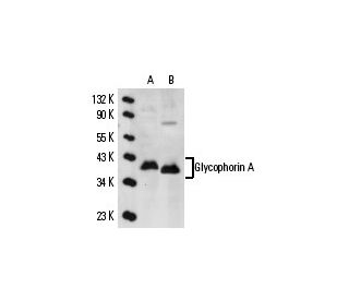
Glycophorin A Antibody (R10) is a mouse monoclonal IgG1 kappa light chain antibody that detects Glycophorin A of human origin by western blotting (WB), immunoprecipitation (IP), immunofluorescence (IF), immunohistochemistry with paraffin-embedded sections (IHCP), and flow cytometry (FCM). Anti-Glycophorin A antibody (R10) is available in both non-conjugated and various conjugated forms, including agarose, horseradish peroxidase (HRP), phycoerythrin (PE), fluorescein isothiocyanate (FITC), and multiple Alexa Fluor® conjugates. Glycophorin A is a crucial sialoglycoprotein located on the surface of human erythrocytes, playing a significant role in maintaining red blood cell structural integrity and facilitating immune system interaction. Glycophorin A spans the membrane once, presenting an amino-terminal end to the extracellular environment, essential for blood group antigenicity function. The genetic diversity of glycophorin surface antigens, including Glycophorin A, B, and C, determines blood group phenotype, making these proteins vital for blood transfusion compatibility and understanding hemolytic diseases. The Glycophorin A gene, located on chromosome 4q31.21, consists of seven exons and exhibits high homology with Glycophorin B, encoding a 91 amino acid protein. Understanding Glycophorin A structure and function remains crucial for clinical applications and erythrocyte biology research, providing insights into blood group antigen expression and potential therapeutic targets for blood-related disorders.
Alexa Fluor® is a trademark of Molecular Probes Inc., OR., USA
LI-COR® and Odyssey® are registered trademarks of LI-COR Biosciences
Glycophorin A Antibody (R10) References:
- Immunohistochemical identification of erythroid precursors in paraffin embedded bone marrow sections: spectrin is a superior marker to glycophorin. | Sadahira, Y., et al. 1999. J Clin Pathol. 52: 919-21. PMID: 10711257
- In vivo detection of hetero-association of glycophorin-A and its mutants within the membrane. | Gerber, D. and Shai, Y. 2001. J Biol Chem. 276: 31229-32. PMID: 11402026
- Distinct regions of human glycophorin A enhance human red cell anion exchanger (band 3; AE1) transport function and surface trafficking. | Young, MT. and Tanner, MJ. 2003. J Biol Chem. 278: 32954-61. PMID: 12813056
- Glycophorin A requirement for expression of O-linked antigens on the erythrocyte membrane. | Arimitsu, N., et al. 2003. Genes Cells. 8: 769-77. PMID: 12940824
- Altered structure and anion transport properties of band 3 (AE1, SLC4A1) in human red cells lacking glycophorin A. | Bruce, LJ., et al. 2004. J Biol Chem. 279: 2414-20. PMID: 14604989
- Structural adaptation of the glycophorin A transmembrane homodimer to D-amino acid modifications. | Gerber, D., et al. 2004. J Mol Biol. 339: 243-50. PMID: 15123435
- Complex interactions at the helix-helix interface stabilize the glycophorin A transmembrane dimer. | Doura, AK. and Fleming, KG. 2004. J Mol Biol. 343: 1487-97. PMID: 15491626
- Co-transfection of murine NXPE2 and murine glycophorin A confers reactivity with Ter-119. | Keele, GR., et al. 2024. Haematologica. 109: 3755-3759. PMID: 39021224
- Glycophorin A as a cell surface marker of early erythroid differentiation in acute leukemia. | Andersson, LC., et al. 1979. Int J Cancer. 24: 717-20. PMID: 397196
- Glycophorin A expression in malignant hematopoiesis. | Liszka, K., et al. 1983. Am J Hematol. 15: 219-26. PMID: 6638008
- Cell surface antigen expression in human erythroid progenitors: erythroid and megakaryocytic markers. | Nakahata, T. and Okumura, N. 1994. Leuk Lymphoma. 13: 401-9. PMID: 8069185
- Differentiation between bruises and putrefactive discolorations of the skin by immunological analysis of glycophorin A. | Kibayashi, K., et al. 1993. Forensic Sci Int. 61: 111-7. PMID: 8307520
Ordering Information
| Product Name | Catalog # | UNIT | Price | Qty | FAVORITES | |
Glycophorin A Antibody (R10) | sc-53905 | 200 µg/ml | $322.00 | |||
Glycophorin A Antibody (R10): m-IgG Fc BP-HRP Bundle | sc-528587 | 200 µg Ab; 10 µg BP | $361.00 | |||
Glycophorin A Antibody (R10): m-IgGκ BP-HRP Bundle | sc-520999 | 200 µg Ab, 40 µg BP | $361.00 | |||
Glycophorin A Antibody (R10): m-IgG1 BP-HRP Bundle | sc-543011 | 200 µg Ab; 20 µg BP | $361.00 | |||
Glycophorin A Antibody (R10) AC | sc-53905 AC | 500 µg/ml, 25% agarose | $424.00 | |||
Glycophorin A Antibody (R10) HRP | sc-53905 HRP | 200 µg/ml | $322.00 | |||
Glycophorin A Antibody (R10) FITC | sc-53905 FITC | 200 µg/ml | $336.00 | |||
Glycophorin A Antibody (R10) PE | sc-53905 PE | 200 µg/ml | $349.00 | |||
Glycophorin A Antibody (R10) Alexa Fluor® 488 | sc-53905 AF488 | 200 µg/ml | $364.00 | |||
Glycophorin A Antibody (R10) Alexa Fluor® 546 | sc-53905 AF546 | 200 µg/ml | $364.00 | |||
Glycophorin A Antibody (R10) Alexa Fluor® 594 | sc-53905 AF594 | 200 µg/ml | $364.00 | |||
Glycophorin A Antibody (R10) Alexa Fluor® 647 | sc-53905 AF647 | 200 µg/ml | $364.00 | |||
Glycophorin A Antibody (R10) Alexa Fluor® 680 | sc-53905 AF680 | 200 µg/ml | $364.00 | |||
Glycophorin A Antibody (R10) Alexa Fluor® 790 | sc-53905 AF790 | 200 µg/ml | $364.00 |