")
: sc-7385. Western blot analysis of WT1 expression in non-transfected: sc-117752 (A) and truncated human WT1 transfected: sc-117178 (B) 293T whole cell lysates.")
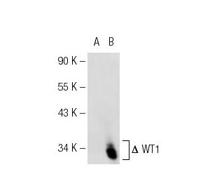
: sc-7385. Western blot analysis of WT1 expression in K-562 whole cell lysate.")
WT1 Antibody (F-6): sc-7385
- WT1 Antibody (F-6) is a mouse monoclonal IgG1 WT1 antibody, cited in 154 publications, provided at 200 µg/ml
- epitope corresponding to a domain 180 amino acids in length mapping near the N-terminus of WT1 of human origin
- WT1 Antibody (F-6) is recommended for detection of WT1 of mouse, rat and human origin by WB, IP and IF
- WT1 Antibody (F-6) is available conjugated to agarose for IP
- WT1 Antibody (F-6) is available conjugated to Alexa Fluor® 546 or Alexa Fluor® 594 for WB (RGB), IF, IHC(P) and FCM
- also available conjugated to Alexa Fluor® 680 or Alexa Fluor® 790 for WB (NIR), IF and FCM; for use with Near-Infrared (NIR) detection systems, such as LI-COR®Odyssey®, iBright™ FL1000, FluorChem™, Typhoon, Azure and other comparable systems
- TransCruz reagent for Gel Supershift and ChIP applications (sc-7385 X, 200 µg/0.1 ml)
- m-IgG1 BP-HRP is the preferred secondary detection reagent for WT1 Antibody (F-6) for WB applications. This reagent is now offered in a bundle with WT1 Antibody (F-6) (see ordering information below).

QUICK LINKS
SEE ALSO...
PKC ζ Antibody (H-1) is a mouse monoclonal IgG2a antibody that targets an epitope mapping between amino acids 573-592 at the C-terminus of rat-origin Protein Kinase C zeta (PKC ζ), also known as PRKCZ or nPKC-ζ. PKC ζ monoclonal antibody (H-1) reacts with mouse, rat, and human species and is suitable for applications including western blotting (WB), immunoprecipitation (IP), immunofluorescence (IF), immunohistochemistry on paraffin-embedded sections (IHCP), and enzyme-linked immunosorbent assay (ELISA). PKC ζ is an atypical member of the protein kinase C family, playing a crucial role in various cellular processes such as cell growth, differentiation, survival, gene expression, and membrane function. Unlike conventional PKCs, PKC ζ activation is independent of calcium and diacylglycerol (DAG), allowing unique function within signaling pathways. PKC ζ is involved in the regulation of NF-κB activation, insulin signaling, cell polarity, and the PI3K/Akt pathway, making PKC ζ significant in understanding diseases like diabetes, cancer, and immune disorders. PKC ζ involvement in cell survival and proliferation underscores importance as a potential therapeutic target. Furthermore, distinct activation mechanisms make PKC ζ an intriguing subject in signal transduction research, potentially revealing new insights into cellular communication pathways. PKC ζ monoclonal antibody (H-1) enables researchers to effectively study expression and function in various biological contexts, aiding in the development of novel treatments.
Alexa Fluor® is a trademark of Molecular Probes Inc., OR., USA
LI-COR® and Odyssey® are registered trademarks of LI-COR Biosciences
WT1 Antibody (F-6) References:
- Zinc finger point mutations within the WT1 gene in Wilms tumor patients. | Little, MH., et al. 1992. Proc Natl Acad Sci U S A. 89: 4791-5. PMID: 1317572
- Repression of the insulin-like growth factor II gene by the Wilms tumor suppressor WT1. | Drummond, IA., et al. 1992. Science. 257: 674-8. PMID: 1323141
- The Wilms' tumor gene product, WT1, represses transcription of the platelet-derived growth factor A-chain gene. | Wang, ZY., et al. 1992. J Biol Chem. 267: 21999-2002. PMID: 1429549
- Characterization of the zinc finger protein encoded by the WT1 Wilms' tumor locus. | Morris, JF., et al. 1991. Oncogene. 6: 2339-48. PMID: 1662794
- A tumor suppressor and oncogene: the WT1 story. | Yang, L., et al. 2007. Leukemia. 21: 868-76. PMID: 17361230
- Isolation and characterization of a zinc finger polypeptide gene at the human chromosome 11 Wilms' tumor locus. | Call, KM., et al. 1990. Cell. 60: 509-20. PMID: 2154335
- Homozygous deletion in Wilms tumours of a zinc-finger gene identified by chromosome jumping. | Gessler, M., et al. 1990. Nature. 343: 774-8. PMID: 2154702
- Introduction of a normal human chromosome 11 into a Wilms' tumor cell line controls its tumorigenic expression. | Weissman, BE., et al. 1987. Science. 236: 175-80. PMID: 3031816
- Genetics of Wilms' tumor. | Matsunaga, E. 1981. Hum Genet. 57: 231-46. PMID: 6265341
Ordering Information
| Product Name | Catalog # | UNIT | Price | Qty | FAVORITES | |
WT1 Antibody (F-6) | sc-7385 | 200 µg/ml | $322.00 | |||
WT1 Antibody (F-6): m-IgG1 BP-HRP Bundle | sc-551910 | 200 µg Ab; 20 µg BP | $361.00 | |||
WT1 Antibody (F-6) X | sc-7385 X | 200 µg/0.1 ml | $322.00 | |||
WT1 Antibody (F-6) AC | sc-7385 AC | 500 µg/ml, 25% agarose | $424.00 | |||
WT1 Antibody (F-6) Alexa Fluor® 546 | sc-7385 AF546 | 200 µg/ml | $364.00 | |||
WT1 Antibody (F-6) Alexa Fluor® 594 | sc-7385 AF594 | 200 µg/ml | $364.00 | |||
WT1 Antibody (F-6) Alexa Fluor® 680 | sc-7385 AF680 | 200 µg/ml | $364.00 | |||
WT1 Antibody (F-6) Alexa Fluor® 790 | sc-7385 AF790 | 200 µg/ml | $364.00 |