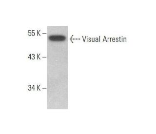
Date published: 2025-9-30

1-800-457-3801

SCBT Portrait Logo
Seach Input

Visual Arrestin Antibody (C-1): sc-271159

5.0(1)
Write a reviewAsk a question

Datasheets
  • Visual Arrestin Antibody (C-1) is a mouse monoclonal IgG1 κ, cited in 1 publications, provided at 200 µg/ml
  • raised against a peptide mapping within an internal region of Visual Arrestin of human origin
  • recommended for detection of Visual Arrestin of mouse, rat and human origin by WB, IP, IF, IHC(P) and ELISA; also reactive with additional species, including and equine, canine and bovine
  • m-IgG Fc BP-HRP, m-IgG1 BP-HRP and m-IgGκ BP-HRP are the preferred secondary detection reagents for Visual Arrestin Antibody (C-1) for WB and IHC(P) applications. These reagents are now offered in bundles with Visual Arrestin Antibody (C-1) (see ordering information below).

QUICK LINKS

SEE ALSO...

Visual Arrestin Antibody (C-1) is a mouse monoclonal IgG1 kappa light chain antibody designed to detect Visual Arrestin, also known as Arrestin 1, retinal S-antigen, or S-Arrestin, in multiple species including human, mouse, rat, equine, canine, and bovine. Anti-Visual Arrestin antibody (C-1) is raised against a peptide within an internal region of the human Visual Arrestin protein, enabling reliable detection in applications such as western blotting (WB), immunoprecipitation (IP), immunofluorescence (IF), immunohistochemistry with paraffin-embedded sections (IHCP), and enzyme-linked immunosorbent assay (ELISA). Visual Arrestin plays a crucial role in the phototransduction pathway by binding to phosphorylated rhodopsin, thereby terminating the light signal and preventing continuous activation of G protein-coupled receptors. Additionally, Visual Arrestin interacts with G protein-coupled receptor kinases (GRKs) to facilitate receptor desensitization and promote receptor internalization through clathrin-mediated endocytosis. These interactions are essential for maintaining precise control over visual signal transduction, ensuring accurate visual perception and preventing signal desensitization. Visual Arrestin is predominantly localized in the outer segments of retinal photoreceptor cells and the pineal gland, where Visual Arrestin contributes to the regulation of circadian rhythms. The strategic cellular localization and interaction with key proteins like rhodopsin and GRKs highlight Visual Arrestin′s importance in both vision and overall physiological regulation. Furthermore, Visual Arrestin serves as a major pathogenic autoantigen in inflammatory eye diseases such as uveoretinitis and Oguchi disease, a rare form of night blindness, underscoring Visual Arrestin′s significance in ocular health and disease. Visual Arrestin (C-1) antibody facilitates comprehensive studies of this protein′s function and interactions, supporting research into visual signal transduction, retinal disorders, and autoimmune conditions.

For Research Use Only. Not Intended for Diagnostic or Therapeutic Use.

Ordering Information

Product NameCatalog #UNITPriceQtyFAVORITES

Visual Arrestin Antibody (C-1)

sc-271159
200 µg/ml
$316.00

Visual Arrestin Antibody (C-1): m-IgG Fc BP-HRP Bundle

sc-537575
200 µg Ab; 10 µg BP
$354.00

Visual Arrestin Antibody (C-1): m-IgGκ BP-HRP Bundle

sc-534879
200 µg Ab; 40 µg BP
$354.00

Visual Arrestin Antibody (C-1): m-IgG1 BP-HRP Bundle

sc-545239
200 µg Ab; 20 µg BP
$354.00

Visual Arrestin (C-1) Neutralizing Peptide

sc-271159 P
100 µg/0.5 ml
$68.00

What application is the blocking peptide sc-271159 P appropriate for?

Asked by: Professor Griffin
Thank you for your question. The blocking peptide is intended for use as a negative control, by pre-adsorbing the mouse monoclonal antibody against the antigen. For full protocol details, please contact our Technical Services Department or view our online protocol here: https://www.scbt.com/scbt/resources/protocols/peptide-neutralization
Answered by: Technical Support
Date published: 2017-02-25
  • y_2025, m_9, d_24, h_6CST
  • bvseo_bulk, prod_bvqa, vn_bulk_3.0.42
  • cp_1, bvpage1
  • co_hasquestionsanswers, tq_1
  • loc_en_US, sid_271159, prod, sort_[SortEntry(order=LAST_APPROVED_ANSWER_SUBMISSION_TIME, direction=DESCENDING)]
  • clientName_scbt
  • bvseo_sdk, java_sdk, bvseo-4.0.0
  • CLOUD, getContent, 101ms
  • QUESTIONS, PRODUCT
Rated 5 out of 5 by from Great Western Blot data of Visual ArrestinGreat Western Blot data of Visual Arrestin expression in non-transfected and human Visual Arrestin transfected 293 whole cell lysates and rat eye tissue extract . -SCBT QC
Date published: 2014-04-25
  • y_2025, m_9, d_24, h_6
  • bvseo_bulk, prod_bvrr, vn_bulk_3.0.42
  • cp_1, bvpage1
  • co_hasreviews, tv_0, tr_1
  • loc_en_US, sid_271159, prod, sort_[SortEntry(order=SUBMISSION_TIME, direction=DESCENDING)]
  • clientName_scbt
  • bvseo_sdk, java_sdk, bvseo-4.0.0
  • CLOUD, getReviews, 16ms
  • REVIEWS, PRODUCT
Visual Arrestin Antibody (C-1) is rated 5.0 out of 5 by 1.
  • y_2025, m_9, d_24, h_6
  • bvseo_bulk, prod_bvrr, vn_bulk_3.0.42
  • cp_1, bvpage1
  • co_hasreviews, tv_0, tr_1
  • loc_en_US, sid_271159, prod, sort_[SortEntry(order=SUBMISSION_TIME, direction=DESCENDING)]
  • clientName_scbt
  • bvseo_sdk, java_sdk, bvseo-4.0.0
  • CLOUD, getAggregateRating, 96ms
  • REVIEWS, PRODUCT