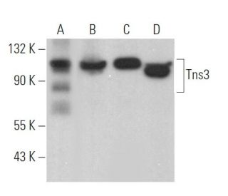
Date published: 2026-4-24

1-800-457-3801

SCBT Portrait Logo
Seach Input

Tns3 Antibody (C-2): sc-376367

4.8(5)
Write a reviewAsk a question

Datasheets
  • Tns3 Antibody (C-2) is a mouse monoclonal IgG2a κ Tns3 antibody, cited in 2 publications, provided at 200 µg/ml
  • raised against amino acids 721-1020 mapping within an internal region of Tns3 of human origin
  • Tns3 Antibody (C-2) is recommended for detection of Tns3 of mouse, rat and human origin by WB, IP, IF, IHC(P) and ELISA
  • Anti-Tns3 Antibody (C-2) is available conjugated to agarose for IP; HRP for WB, IHC(P) and ELISA; and to either phycoerythrin or FITC for IF, IHC(P) and FCM
  • also available conjugated to Alexa Fluor® 488, Alexa Fluor® 546, Alexa Fluor® 594 or Alexa Fluor® 647 for WB (RGB), IF, IHC(P) and FCM, and for use with RGB fluorescent imaging systems, such as iBright™ FL1000, FluorChem™, Typhoon, Azure and other comparable systems
  • also available conjugated to Alexa Fluor® 680 or Alexa Fluor® 790 for WB (NIR), IF and FCM; for use with Near-Infrared (NIR) detection systems, such as LI-COR®Odyssey®, iBright™ FL1000, FluorChem™, Typhoon, Azure and other comparable systems
  • m-IgG Fc BP-HRP, m-IgG2a BP-HRP and m-IgGκ BP-HRP are the preferred secondary detection reagents for Tns3 Antibody (C-2) for WB and IHC(P) applications. These reagents are now offered in bundles with Tns3 Antibody (C-2) (see ordering information below).
Gene Editing Promo Banner

QUICK LINKS

SEE ALSO...

Tns3 Antibody (C-2) is a mouse monoclonal IgG2a kappa light chain antibody that detects Tns3 protein of mouse, rat, and human origin by western blotting (WB), immunoprecipitation (IP), immunofluorescence (IF), immunohistochemistry, and enzyme-linked immunosorbent assay (ELISA). Tns3 (C-2) antibody is available in both non-conjugated and various conjugated forms, including agarose, horseradish peroxidase (HRP), phycoerythrin (PE), fluorescein isothiocyanate (FITC), and multiple Alexa Fluor® conjugates. Tns3, also known as TEM6 or TENS1, plays a crucial role in cellular structure maintenance by anchoring actin filaments at focal adhesions, which is vital for cell adhesion and signaling. Tns3 is predominantly localized at focal adhesions of the plasma membrane and is essential for proper growth and development, as evidenced by growth retardation and mortality observed in Tns3-deficient mice. Tns3 shows high expression in thyroid and placenta, while maintaining presence in heart, liver, brain, prostate, pancreas, kidney, lung, skeletal muscle, and white blood cells. A Src homology 2 (SH2) domain in Tns3 suggests involvement in signal transduction pathways, linking cytoskeleton to cellular signaling mechanisms. These diverse functions and interactions of Tns3 highlight significance in both structural integrity and cellular communication, making anti-Tns3 antibody (C-2) a valuable tool for researchers studying these critical biological processes.

For Research Use Only. Not Intended for Diagnostic or Therapeutic Use.

Alexa Fluor® is a trademark of Molecular Probes Inc., OR., USA

LI-COR® and Odyssey® are registered trademarks of LI-COR Biosciences

Tns3 Antibody (C-2) References:

  1. Epidermal growth factor modulates tyrosine phosphorylation of a novel tensin family member, tensin3.  |  Cui, Y., et al. 2004. Mol Cancer Res. 2: 225-32. PMID: 15140944
  2. Inactivation of tensin3 in mice results in growth retardation and postnatal lethality.  |  Chiang, MK., et al. 2005. Dev Biol. 279: 368-77. PMID: 15733665
  3. Tensin3 is a novel thyroid-specific gene.  |  Maeda, I., et al. 2006. J Mol Endocrinol. 36: R1-8. PMID: 16461921
  4. Tensin: a potential link between the cytoskeleton and signal transduction.  |  Lo, SH., et al. 1994. Bioessays. 16: 817-23. PMID: 7840759
  5. Molecular cloning, expression, and mapping of the high affinity actin-capping domain of chicken cardiac tensin.  |  Chuang, JZ., et al. 1995. J Cell Biol. 128: 1095-109. PMID: 7896874
  6. Interactions of tensin with actin and identification of its three distinct actin-binding domains.  |  Lo, SH., et al. 1994. J Cell Biol. 125: 1067-75. PMID: 8195290
  7. Cell spreading on extracellular matrix proteins induces tyrosine phosphorylation of tensin.  |  Bockholt, SM. and Burridge, K. 1993. J Biol Chem. 268: 14565-7. PMID: 8325835
  8. The N-terminal domains of tensin and auxilin are phosphatase homologues.  |  Haynie, DT. and Ponting, CP. 1996. Protein Sci. 5: 2643-6. PMID: 8976573

Ordering Information

Product NameCatalog #UNITPriceQtyFAVORITES

Tns3 Antibody (C-2)

sc-376367
200 µg/ml
$322.00

Tns3 Antibody (C-2): m-IgG Fc BP-HRP Bundle

sc-529859
200 µg Ab; 10 µg BP
$361.00

Tns3 Antibody (C-2): m-IgGκ BP-HRP Bundle

sc-522929
200 µg Ab, 40 µg BP
$361.00

Tns3 Antibody (C-2): m-IgG2a BP-HRP Bundle

sc-547392
200 µg Ab; 10 µg BP
$361.00

Tns3 Antibody (C-2) AC

sc-376367 AC
500 µg/ml, 25% agarose
$424.00

Tns3 Antibody (C-2) HRP

sc-376367 HRP
200 µg/ml
$322.00

Tns3 Antibody (C-2) FITC

sc-376367 FITC
200 µg/ml
$336.00

Tns3 Antibody (C-2) PE

sc-376367 PE
200 µg/ml
$349.00

Tns3 Antibody (C-2) Alexa Fluor® 488

sc-376367 AF488
200 µg/ml
$364.00

Tns3 Antibody (C-2) Alexa Fluor® 546

sc-376367 AF546
200 µg/ml
$364.00

Tns3 Antibody (C-2) Alexa Fluor® 594

sc-376367 AF594
200 µg/ml
$364.00

Tns3 Antibody (C-2) Alexa Fluor® 647

sc-376367 AF647
200 µg/ml
$364.00

Tns3 Antibody (C-2) Alexa Fluor® 680

sc-376367 AF680
200 µg/ml
$364.00

Tns3 Antibody (C-2) Alexa Fluor® 790

sc-376367 AF790
200 µg/ml
$364.00

Dear, I already ordered for this antibody, and I want to know the concentration or dilution for the immunostaining you recommend ? thank you in advance Hatem

Asked by: Hatem
Thank you for your question. The recommended starting dilution for immunostaining is 1:50 with a dilution range of 1:50-1:500. This information, along with other technical specifications, can be found in the “datasheet” link at the top of the product page. If you have any further questions or concerns, please contact our Technical Service department by phone at Toll Free: +49 6221 4503 0, by email at: europe@scbt.com or by live chat directly on our website, www.scbt.com
Answered by: Tech Service
Date published: 2017-04-21

I am using Tns3 (C-2): sc-376367 monoclonal antibody to stain paraffin embedded tissue. Do you recommend antigen retrieval when using this antibody?

Asked by: Professor Griffin
We recommend performing antigen retrieval with sodium citrate buffer (pH 6) and heat. The full protocol can be found here: https://www.scbt.com/scbt/resources/protocols/immunoperoxidase-staining
Answered by: Technical Support
Date published: 2017-02-24
  • y_2026, m_4, d_23, h_7CST
  • bvseo_bulk, prod_bvqa, vn_bulk_3.0.43
  • cp_1, bvpage1
  • co_hasquestionsanswers, tq_2
  • loc_en_US, sid_376367, prod, sort_[SortEntry(order=LAST_APPROVED_ANSWER_SUBMISSION_TIME, direction=DESCENDING)]
  • clientName_scbt
  • bvseo_sdk, java_sdk, bvseo-4.0.0
  • CLOUD, getContent, 119ms
  • QUESTIONS, PRODUCT
Rated 5 out of 5 by from Works well for WB and IFThe antibody gives strong and clean signal in WB and was also working well in IF.
Date published: 2020-10-06
Rated 4 out of 5 by from Good results in IFI received a sample product a couple of months ago and tested it on MDA-MB-231 breast cancer cells, plated on collagen. This is the best Ab for TNS3 that I have used so far. It clearly labels adhesion site and this is strongly abolished upon transfection of an siRNA targeting TNS3. There is some cytosolic staining (which is reduced upon the KD) and some nuclear staining that seems unspecific, as it is still present in the KD cells (not visible in the images below, as they are taken with a confocal microscope focusing on the adhesion plane). Despite this, I think it is a very good antibody to assess TNS3 localisation in adhesive complexes.
Date published: 2018-02-01
Rated 5 out of 5 by from Good all aroundWell established clone for use in Western Blot, but also performs well in IF and IHC with a variety of fixation methods.
Date published: 2017-02-06
Rated 5 out of 5 by from Superb immunofluorescence focal adhesionsSuperb immunofluorescence focal adhesions and cytoplasmic staining in formalin-fixed HepG2 cells. -SCBT QC
Date published: 2015-03-26
  • y_2026, m_4, d_23, h_7
  • bvseo_bulk, prod_bvrr, vn_bulk_3.0.43
  • cp_1, bvpage1
  • co_hasreviews, tv_1, tr_4
  • loc_en_US, sid_376367, prod, sort_[SortEntry(order=SUBMISSION_TIME, direction=DESCENDING)]
  • clientName_scbt
  • bvseo_sdk, java_sdk, bvseo-4.0.0
  • CLOUD, getReviews, 15ms
  • REVIEWS, PRODUCT
Tns3 Antibody (C-2) is rated 4.8 out of 5 by 5.
  • y_2026, m_4, d_23, h_7
  • bvseo_bulk, prod_bvrr, vn_bulk_3.0.43
  • cp_1, bvpage1
  • co_hasreviews, tv_1, tr_4
  • loc_en_US, sid_376367, prod, sort_[SortEntry(order=SUBMISSION_TIME, direction=DESCENDING)]
  • clientName_scbt
  • bvseo_sdk, java_sdk, bvseo-4.0.0
  • CLOUD, getAggregateRating, 105ms
  • REVIEWS, PRODUCT