")
Rb Anticorpo (H-2): sc-74570
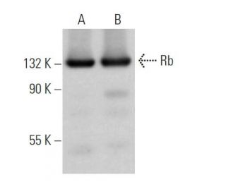
- Rb Anticorpo H-2é um anticorpo monoclonal produzido em camundongo IgG2a κ, citado em 7 publicações, fornecido em 200 µg/ml
- anti-aminioácidos 769-921 mapeamento no C-terminus de Rb de mouse origem
- recomendado para a detecção de Rb de mouse, rat e human origem em
- See Rb (IF8): sc-102 for additional antibody conjugates, including AC, HRP, FITC, PE, Alexa Fluor® 488, 594, 647, 680 and 790.
- m-IgG Fc BP-HRP e 2a BP-HRP">m-IgG2a BP-HRP são os reagentes de deteção secundários preferidos para Rb Antibody (H-2) para aplicações WB. Estes reagentes são agora oferecidos em conjuntos com Rb Antibody (H-2)(ver informações de encomenda abaixo).

LINKS RÁPIDOS
VEJA TAMBÉM
O câncer pediátrico retinoblastoma e a formação de outros tumores humanos podem ser atribuídos a mutações no gene supressor de tumor retinoblastoma. O produto do gene supressor de tumor do retinoblastoma, conhecido como Rb ou pRb, regula a diferenciação, a apoptose e o controle do ciclo celular coordenando o ciclo celular, em G1/S, com o mecanismo de transcrição que inclui a família E2F heterodimérica. Durante G1, a fosforilação de Rb mediada por quinase dependente de ciclina D (D1, D2, D3) em Ser 795 marca a conversão de Rb de um estado hipofosforilado e repressivo de transcrição para um estado inativo e fosforilado, que pode ser mantido durante a mitose por fosforilação diferencial de até 16 resíduos putativos de serina ou treonina, incluindo Ser 249/Thr 252, Thr 373, Thr 356, Ser 780, Ser 807/Ser 811 e Thr 821/Thr 826. A Rb hipofosforilada reprime a transcrição de genes que controlam o ciclo celular por meio de interações diretas proteína-proteína, ligando e inativando os promotores de fatores de transcrição e por meio do recrutamento de histona desacetilase, que desacetilam as regiões do promotor e aumentam a formação de nucleossomos, mascarando assim os elementos cis que aumentam a transcrição.
Informacoes sobre ordens
| Nome do Produto | Numero de Catalogo | UNID | Preco | Qde | FAVORITOS | |
Rb Anticorpo (H-2) | sc-74570 | 200 µg/ml | $322.00 | |||
Pacote do Rb (H-2): m-IgG Fc BP-HRP | sc-539548 | 200 µg Ab; 10 µg BP | $361.00 | |||
Pacote do Rb (H-2): m-IgG2a BP-HRP | sc-546731 | 200 µg Ab; 10 µg BP | $361.00 |