")
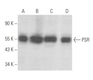
PSR Anticorpo (H-7): sc-28348
- PSR Anticorpo H-7é um anticorpo monoclonal produzido em camundongo IgG1 κ PSR anticorpo, citado em 32 publicações, fornecido em 200 µg/ml
- Produzido contra aminoácidos 1-300 de PSR de human origem
- PSR Anticorpo (H-7) é recomendado para a detecção de PSR de e human origem em WB, IP, IF, IHC(P) e ELISA
- Anticorpo anti-PSR O anticorpo anti-PSR (H-7) está disponível conjugado com agarose para IP; HRP para WB, IHC(P) e ELISA; e com fioeritrina ou FITC para IF, IHC(P) e FCM
- também disponível conjugado com Alexa Fluor® 488, Alexa Fluor® 546, Alexa Fluor® 594 ou Alexa Fluor® 647 para WB (RGB), IF, IHC(P) e FCM, e para utilização com sistemas de imagiologia fluorescente RGB, tais como iBright™ FL1000, FluorChem™, Typhoon, Azure e outros sistemas comparáveis
- também disponível conjugado com Alexa Fluor® 680 ou Alexa Fluor® 790 para WB (NIR), IF e FCM; para utilização com sistemas de deteção de infravermelhos próximos (NIR), tais como LI-COR®Odyssey®, iBright™ FL1000, FluorChem™, Typhoon, Azure e outros sistemas comparáveis
- O m-IgG Fc BP-HRP e o m-IgGκ BP-HRP são os reagentes de deteção secundários preferidos para o anticorpo PSR (H-7) para aplicações WB e IHC(P). Estes reagentes são agora disponibilizados em conjuntos com o anticorpo PSR (H-7)(ver informações de encomenda abaixo).

LINKS RÁPIDOS
VEJA TAMBÉM
O Anticorpo PSR (H-7) é um anticorpo monoclonal de cadeia leve IgG1 kappa de camundongo que detecta a proteína PSR de origem humana por western blotting (WB), imunoprecipitação (IP), imunofluorescência (IF), imunohistoquímica com secções incluídas em parafina (IHCP) e ensaio de imunoabsorção enzimática (ELISA). O anticorpo anti-PSR (H-7) está disponível tanto em formas não conjugadas como em várias formas conjugadas, incluindo agarose, peroxidase de rábano (HRP), ficoeritrina (PE), isotiocianato de fluoresceína (FITC) e vários conjugados Alexa Fluor®. O recetor de fosfatidilserina (PSR) desempenha um papel crucial no reconhecimento e eliminação de células apoptóticas, uma vez que o PSR se liga especificamente à fosfatidilserina, que fica exposta na membrana externa das células em processo de apoptose. Esta interação é vital para a fagocitose das células moribundas pelos macrófagos e células dendríticas, prevenindo assim a inflamação e mantendo a homeostasia dos tecidos. A PSR é predominantemente expressa na superfície de macrófagos, fibroblastos e células epiteliais, com altos níveis de expressão observados nos tecidos do coração, do músculo esquelético e dos rins. Além disso, a PSR é extensivamente glicosilada, o que pode influenciar as propriedades de ligação e as interações com outras proteínas. A conservação da PSR em todas as espécies, como evidenciado pela homologia significativa com proteínas encontradas em Caenorhabditis elegans e Drosophila melanogaster, sublinha o papel fundamental da PSR nos processos celulares ao longo da evolução.
Alexa Fluor® é uma marca comercial da Molecular Probes Inc., OR., EUA
LI-COR® e Odyssey® são marcas registadas da LI-COR Biosciences
Referencias do PSR Anticorpo (H-7):
- O papel da fosfatidilserina no reconhecimento de células apoptóticas pelos fagócitos. | Fadok, VA., et al. 1998. Cell Death Differ. 5: 551-62. PMID: 10200509
- A exposição da fosfatidilserina é uma caraterística geral da fagocitose de linfócitos apoptóticos pelos macrófagos. | Krahling, S., et al. 1999. Cell Death Differ. 6: 183-9. PMID: 10200565
- Necessidade de croquemort na fagocitose de células apoptóticas em Drosophila. | Franc, NC., et al. 1999. Science. 284: 1991-4. PMID: 10373118
- Apoptose. Desaparecido mas não esquecido. | Green, DR. and Beere, HM. 2000. Nature. 405: 28-9. PMID: 10811203
- Um recetor para a eliminação específica de fosfatidilserina de células apoptóticas. | Fadok, VA., et al. 2000. Nature. 405: 85-90. PMID: 10811223
- Localização nuclear da proteína recetora da fosfatidilserina através de múltiplos sinais de localização nuclear. | Cui, P., et al. 2004. Exp Cell Res. 293: 154-63. PMID: 14729065
- O recetor da fosfatidilserina coopera com o recetor da lipoproteína de alta densidade no reconhecimento de células apoptóticas pelas células da enfermeira tímica. | Cao, WM., et al. 2004. J Mol Endocrinol. 32: 497-505. PMID: 15072554
- A exposição da fosfatidilserina na superfície dos linfócitos apoptóticos desencadeia o reconhecimento específico e a remoção pelos macrófagos. | Fadok, VA., et al. 1992. J Immunol. 148: 2207-16. PMID: 1545126
- A interação entre a fosfatidilserina e o recetor da fosfatidilserina inibe as respostas imunitárias in vivo. | Hoffmann, PR., et al. 2005. J Immunol. 174: 1393-404. PMID: 15661897
- O recetor de fosfatidilserina Tim-4 é essencial para a manutenção do estado homeostático dos macrófagos peritoneais residentes. | Wong, K., et al. 2010. Proc Natl Acad Sci U S A. 107: 8712-7. PMID: 20421466
- O recrutamento de p85α pelo recetor de fosfatidilserina CD300f medeia a eliminação de células apoptóticas necessária para a supressão da autoimunidade. | Tian, L., et al. 2014. Nat Commun. 5: 3146. PMID: 24477292
- A proteína adaptadora candidata CED-6 promove o engolfamento de células apoptóticas em C. elegans. | Liu, QA. and Hengartner, MO. 1998. Cell. 93: 961-72. PMID: 9635426
Informacoes sobre ordens
| Nome do Produto | Numero de Catalogo | UNID | Preco | Qde | FAVORITOS | |
PSR Anticorpo (H-7) | sc-28348 | 200 µg/ml | $322.00 | |||
Pacote do PSR (H-7): m-IgG Fc BP-HRP | sc-528431 | 200 µg Ab; 10 µg BP | $361.00 | |||
Pacote do PSR (H-7): m-IgGκ BP-HRP | sc-520806 | 200 µg Ab, 40 µg BP | $361.00 | |||
PSR Anticorpo (H-7) AC | sc-28348 AC | 500 µg/ml, 25% agarose | $424.00 | |||
PSR Anticorpo (H-7) HRP | sc-28348 HRP | 200 µg/ml | $322.00 | |||
PSR Anticorpo (H-7) FITC | sc-28348 FITC | 200 µg/ml | $336.00 | |||
PSR Anticorpo (H-7) PE | sc-28348 PE | 200 µg/ml | $349.00 | |||
PSR Anticorpo (H-7) Alexa Fluor® 488 | sc-28348 AF488 | 200 µg/ml | $364.00 | |||
PSR Anticorpo (H-7) Alexa Fluor® 546 | sc-28348 AF546 | 200 µg/ml | $364.00 | |||
PSR Anticorpo (H-7) Alexa Fluor® 594 | sc-28348 AF594 | 200 µg/ml | $364.00 | |||
PSR Anticorpo (H-7) Alexa Fluor® 647 | sc-28348 AF647 | 200 µg/ml | $364.00 | |||
PSR Anticorpo (H-7) Alexa Fluor® 680 | sc-28348 AF680 | 200 µg/ml | $364.00 | |||
PSR Anticorpo (H-7) Alexa Fluor® 790 | sc-28348 AF790 | 200 µg/ml | $364.00 |