")
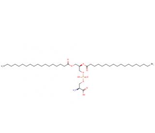

Phosphatidyl-L-serine (CAS 51446-62-9)
QUICK LINKS
Phosphatidyl-L-serine (PS) is a phospholipid component crucial to the structure and function of biological membranes, particularly in the brain and nervous system. Structurally, it consists of two fatty acid chains linked to a glycerol backbone, with one of the hydroxyl groups of glycerol esterified to phosphoric acid, which is further linked to the amino acid serine. This configuration imparts specific biophysical properties to membranes, including fluidity and phase behavior, which are essential for proper cellular function. In research contexts, PS is often studied for its role in signaling pathways associated with apoptosis (programmed cell death) and cellular signaling. The presence of PS on the inner leaflet of the cell membrane′s bilayer is pivotal in maintaining cellular integrity and signaling cascades. However, during apoptosis, PS is translocated to the outer leaflet, where it serves as a signal for phagocytes to engulf apoptotic cells, a process vital for tissue homeostasis and immune response. In scientific studies, PS has been used as a tool to understand membrane asymmetry and the mechanisms underlying apoptosis, without therapeutic intent. Researchers utilize it in a variety of experimental setups, including artificial membrane systems and live cell imaging, to track and manipulate cellular processes under controlled conditions, thereby elucidating the complex interactions of cellular signaling networks. Such studies provide foundational knowledge that can inform broader biological and biochemical insights.
Phosphatidyl-L-serine (CAS 51446-62-9) References
- Interactions of the C2 domain of human factor V with a model membrane. | Mollica, L., et al. 2006. Proteins. 64: 363-75. PMID: 16680712
- Crystal structure of lactadherin C2 domain at 1.7A resolution with mutational and computational analyses of its membrane-binding motif. | Shao, C., et al. 2008. J Biol Chem. 283: 7230-41. PMID: 18160406
- Occurrence of phosphatidyl-D-serine in the rat cerebrum. | Omori, T., et al. 2009. Biochem Biophys Res Commun. 382: 415-8. PMID: 19285036
- Factor VIII C1 domain residues Lys 2092 and Phe 2093 contribute to membrane binding and cofactor activity. | Meems, H., et al. 2009. Blood. 114: 3938-46. PMID: 19687511
- Phosphatidylserine exposure and procoagulant activity in acute promyelocytic leukemia. | Zhou, J., et al. 2010. J Thromb Haemost. 8: 773-82. PMID: 20102487
- Chemotherapy induces enhanced procoagulant activity through phosphatidylserine exposure in acute lymphoblastic leukemia. | Dong, X., et al. 2013. Thromb Res. 132: 614-20. PMID: 24074703
- Lactadherin inhibits secretory phospholipase A2 activity on pre-apoptotic leukemia cells. | Nyegaard, S., et al. 2013. PLoS One. 8: e77143. PMID: 24194865
- Stereochemistry- and concentration-dependent effects of phosphatidylserine enrichment on platelet function. | Meyer, AF., et al. 2017. Biochim Biophys Acta Biomembr. 1859: 1381-1387. PMID: 28472616
- Low-intensity light-induced drug release from a dual delivery system comprising of a drug loaded liposome and a photosensitive conjugate. | Meerovich, I., et al. 2020. J Drug Target. 28: 655-667. PMID: 31886709
- Procoagulant activities of skeletal and cardiac muscle myosin depend on contaminating phospholipid. | Novakovic, VA. and Gilbert, GE. 2020. Blood. 136: 2469-2472. PMID: 32604409
Ordering Information
| Product Name | Catalog # | UNIT | Price | Qty | FAVORITES | |
Phosphatidyl-L-serine, 10 g | sc-507548 | 10 g | $45.00 |