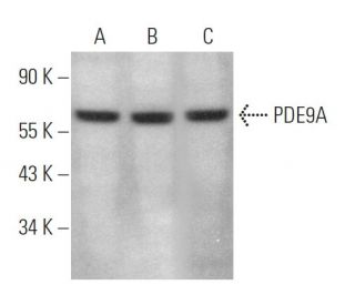
Date published: 2026-3-3

1-800-457-3801

SCBT Portrait Logo
Seach Input

PDE9A Antibody (D-7): sc-376271

5.0(2)
Write a reviewAsk a question

Datasheets
  • PDE9A Antibody (D-7) is a mouse monoclonal IgG1 κ PDE9A antibody provided at 200 µg/ml
  • specific for an epitope mapping between amino acids 225-263 within an internal region of PDE9A of human origin
  • PDE9A Antibody (D-7) is recommended for detection of PDE9A of mouse, rat and human origin by WB, IP, IF, IHC(P) and ELISA; also reactive with additional species, including and canine and porcine
  • Anti-PDE9A Antibody (D-7) is available conjugated to agarose for IP; HRP for WB, IHC(P) and ELISA; and to either phycoerythrin or FITC for IF, IHC(P) and FCM
  • also available conjugated to Alexa Fluor® 488, Alexa Fluor® 546, Alexa Fluor® 594 or Alexa Fluor® 647 for WB (RGB), IF, IHC(P) and FCM, and for use with RGB fluorescent imaging systems, such as iBright™ FL1000, FluorChem™, Typhoon, Azure and other comparable systems
  • also available conjugated to Alexa Fluor® 680 or Alexa Fluor® 790 for WB (NIR), IF and FCM; for use with Near-Infrared (NIR) detection systems, such as LI-COR®Odyssey®, iBright™ FL1000, FluorChem™, Typhoon, Azure and other comparable systems
  • At present, we have not yet completed the identification of the preferred secondary detection reagent(s) for PDE9A Antibody (D-7). This work is in progress.
    Crispr Promo Banner

    QUICK LINKS

    SEE ALSO...

    PDE9A Antibody (D-7) is a mouse monoclonal IgG1 kappa light chain antibody that detects PDE9A of mouse, rat, and human origin by western blotting (WB), immunoprecipitation (IP), immunofluorescence (IF), immunohistochemistry with paraffin-embedded sections (IHCP), and enzyme-linked immunosorbent assay (ELISA). Anti-PDE9A antibody (D-7) is available in both non-conjugated and various conjugated forms, including agarose, horseradish peroxidase (HRP), phycoerythrin (PE), fluorescein isothiocyanate (FITC), and multiple Alexa Fluor® conjugates. Phosphodiesterase 9A (PDE9A) is a 593 amino acid protein that plays a crucial role in signal transduction by regulating intracellular concentration of cyclic nucleotides, particularly exhibiting a high affinity for cyclic guanosine monophosphate (cGMP). This regulation influences various physiological processes, including smooth muscle relaxation, neuronal signaling, and cardiac function. PDE9A is expressed in a range of tissues, with highest levels found in brain, kidney, spleen, colon, heart, and testis, while notably absent in blood. PDE9A consists of an N-terminal regulatory domain and a C-terminal catalytic domain, which contains two potential divalent metal ion binding sites, underscoring enzymatic activity. PDE9A has been implicated in affective bipolar disorder, highlighting significance in neuropsychiatric conditions.

    For Research Use Only. Not Intended for Diagnostic or Therapeutic Use.

    Alexa Fluor® is a trademark of Molecular Probes Inc., OR., USA

    LI-COR® and Odyssey® are registered trademarks of LI-COR Biosciences

    PDE9A Antibody (D-7) References:

    1. Identification and distribution of different mRNA variants produced by differential splicing in the human phosphodiesterase 9A gene.  |  Rentero, C., et al. 2003. Biochem Biophys Res Commun. 301: 686-92. PMID: 12565835
    2. Identification and characterization of a new human type 9 cGMP-specific phosphodiesterase splice variant (PDE9A5). Differential tissue distribution and subcellular localization of PDE9A variants.  |  Wang, P., et al. 2003. Gene. 314: 15-27. PMID: 14527714
    3. Crystal structure of phosphodiesterase 9 shows orientation variation of inhibitor 3-isobutyl-1-methylxanthine binding.  |  Huai, Q., et al. 2004. Proc Natl Acad Sci U S A. 101: 9624-9. PMID: 15210993
    4. Identification and characterization of a novel family of cyclic nucleotide phosphodiesterases.  |  Soderling, SH., et al. 1998. J Biol Chem. 273: 15553-8. PMID: 9624145
    5. Isolation and characterization of PDE9A, a novel human cGMP-specific phosphodiesterase.  |  Fisher, DA., et al. 1998. J Biol Chem. 273: 15559-64. PMID: 9624146
    6. Identification and characterization of a novel cyclic nucleotide phosphodiesterase gene (PDE9A) that maps to 21q22.3: alternative splicing of mRNA transcripts, genomic structure and sequence.  |  Guipponi, M., et al. 1998. Hum Genet. 103: 386-92. PMID: 9856478
    7. Squamous cell carcinoma of the thymus. An analysis of eight cases.  |  Shimosato, Y., et al. 1977. Am J Surg Pathol. 1: 109-21. PMID: 602973

    Ordering Information

    Product NameCatalog #UNITPriceQtyFAVORITES

    PDE9A Antibody (D-7)

    sc-376271
    200 µg/ml
    $322.00

    PDE9A Antibody (D-7) AC

    sc-376271 AC
    500 µg/ml, 25% agarose
    $424.00

    PDE9A Antibody (D-7) HRP

    sc-376271 HRP
    200 µg/ml
    $322.00

    PDE9A Antibody (D-7) FITC

    sc-376271 FITC
    200 µg/ml
    $336.00

    PDE9A Antibody (D-7) PE

    sc-376271 PE
    200 µg/ml
    $349.00

    PDE9A Antibody (D-7) Alexa Fluor® 488

    sc-376271 AF488
    200 µg/ml
    $364.00

    PDE9A Antibody (D-7) Alexa Fluor® 546

    sc-376271 AF546
    200 µg/ml
    $364.00

    PDE9A Antibody (D-7) Alexa Fluor® 594

    sc-376271 AF594
    200 µg/ml
    $364.00

    PDE9A Antibody (D-7) Alexa Fluor® 647

    sc-376271 AF647
    200 µg/ml
    $364.00

    PDE9A Antibody (D-7) Alexa Fluor® 680

    sc-376271 AF680
    200 µg/ml
    $364.00

    PDE9A Antibody (D-7) Alexa Fluor® 790

    sc-376271 AF790
    200 µg/ml
    $364.00

    PDE9A (D-7) Neutralizing Peptide

    sc-376271 P
    100 µg/0.5 ml
    $69.00

    What application is the blocking peptide sc-376271 P appropriate for?

    Asked by: Dr Ninau Qelp
    Thank you for your question. The blocking peptide is intended for use as a negative control, by pre-adsorbing the mouse monoclonal antibody against the antigen. For full protocol details, please contact our Technical Services Department or view our online protocol here: https://www.scbt.com/scbt/resources/protocols/peptide-neutralization
    Answered by: Technical Support
    Date published: 2017-02-24
    • y_2026, m_2, d_26, h_9CST
    • bvseo_bulk, prod_bvqa, vn_bulk_3.0.42
    • cp_1, bvpage1
    • co_hasquestionsanswers, tq_1
    • loc_en_US, sid_376271, prod, sort_[SortEntry(order=LAST_APPROVED_ANSWER_SUBMISSION_TIME, direction=DESCENDING)]
    • clientName_scbt
    • bvseo_sdk, java_sdk, bvseo-4.0.0
    • CLOUD, getContent, 101ms
    • QUESTIONS, PRODUCT
    Rated 5 out of 5 by from Beautiful immunoperoxidase cytoplasmic stainingBeautiful immunoperoxidase cytoplasmic staining in formalin fixed, paraffin-embedded human adrenal gland tissue. -SCBT QC
    Date published: 2015-02-07
    Rated 5 out of 5 by from Satisfactory Western Blot data of PDE9A expressionSatisfactory Western Blot data of PDE9A expression in non-transfected and human PDE9A transfected 293 whole cell lysates. -SCBT QC
    Date published: 2014-07-09
    • y_2026, m_2, d_26, h_9
    • bvseo_bulk, prod_bvrr, vn_bulk_3.0.42
    • cp_1, bvpage1
    • co_hasreviews, tv_0, tr_2
    • loc_en_US, sid_376271, prod, sort_[SortEntry(order=SUBMISSION_TIME, direction=DESCENDING)]
    • clientName_scbt
    • bvseo_sdk, java_sdk, bvseo-4.0.0
    • CLOUD, getReviews, 17ms
    • REVIEWS, PRODUCT
    PDE9A Antibody (D-7) is rated 5.0 out of 5 by 2.
    • y_2026, m_2, d_26, h_9
    • bvseo_bulk, prod_bvrr, vn_bulk_3.0.42
    • cp_1, bvpage1
    • co_hasreviews, tv_0, tr_2
    • loc_en_US, sid_376271, prod, sort_[SortEntry(order=SUBMISSION_TIME, direction=DESCENDING)]
    • clientName_scbt
    • bvseo_sdk, java_sdk, bvseo-4.0.0
    • CLOUD, getAggregateRating, 124ms
    • REVIEWS, PRODUCT