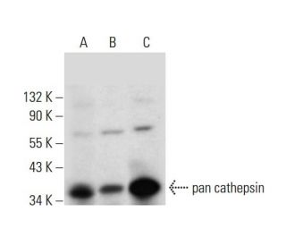
Date published: 2025-11-20

1-800-457-3801

SCBT Portrait Logo
Seach Input

pan cathepsin Antibody (H-1): sc-376803

4.4(5)
Write a reviewAsk a question

Datasheets
  • pan cathepsin Antibody (H-1) is a mouse monoclonal IgG2b κ pan cathepsin antibody, cited in 4 publications, provided at 200 µg/ml
  • specific for an epitope mapping between amino acids 107-145 near the N-terminus of cathepsin of human origin
  • pan cathepsin Antibody (H-1) is recommended for detection of a broad range of cathepsin proteins of mouse, rat and human origin by WB, IP, IF, IHC(P) and ELISA
  • Anti-pan cathepsin Antibody (H-1) is available conjugated to agarose for IP; HRP for WB, IHC(P) and ELISA; and to either phycoerythrin or FITC for IF, IHC(P) and FCM
  • also available conjugated to Alexa Fluor® 488, Alexa Fluor® 546, Alexa Fluor® 594 or Alexa Fluor® 647 for WB (RGB), IF, IHC(P) and FCM, and for use with RGB fluorescent imaging systems, such as iBright™ FL1000, FluorChem™, Typhoon, Azure and other comparable systems
  • also available conjugated to Alexa Fluor® 680 or Alexa Fluor® 790 for WB (NIR), IF and FCM; for use with Near-Infrared (NIR) detection systems, such as LI-COR®Odyssey®, iBright™ FL1000, FluorChem™, Typhoon, Azure and other comparable systems
  • m-IgGκ BP-HRP is the preferred secondary detection reagent for pan cathepsin Antibody (H-1) for WB and IHC(P) applications. This reagent is now offered in a bundle with pan cathepsin Antibody (H-1) (see ordering information below). For additional m-IgGκ BP conjugates see our complete list of Mouse IgG Binding Proteins.

QUICK LINKS

SEE ALSO...

pan cathepsin Antibody (H-1) is a mouse monoclonal IgG2b kappa light chain antibody that detects pan cathepsin protein of mouse, rat, and human origin by western blotting (WB), immunoprecipitation (IP), immunofluorescence (IF), immunohistochemistry with paraffin-embedded sections (IHCP), and enzyme-linked immunosorbent assay (ELISA). pan cathepsin (H-1) antibody is available in both non-conjugated and various conjugated forms, including agarose, horseradish peroxidase (HRP), phycoerythrin (PE), fluorescein isothiocyanate (FITC), and multiple Alexa Fluor® conjugates. The cathepsin family consists of a diverse array of proteolytic enzymes, with cathepsins playing crucial roles in cellular metabolism, including peptide biosynthesis and protein degradation. Notably, cathepsin L, a member of the cysteine protease class, is particularly significant due to its involvement in various physiological processes, such as antigen processing and regulation of apoptosis. Proper functioning of cathepsin L is essential for maintaining cellular homeostasis, and dysregulation of cathepsin L activity has been implicated in several diseases, including cancer and neurodegenerative disorders. Understanding cathepsin L interactions with other proteins, such as cathepsin H, can provide insights into functional mechanisms and potential therapeutic targets.

For Research Use Only. Not Intended for Diagnostic or Therapeutic Use.

Alexa Fluor® is a trademark of Molecular Probes Inc., OR., USA

LI-COR® and Odyssey® are registered trademarks of LI-COR Biosciences

pan cathepsin Antibody (H-1) References:

  1. Molecular cloning and expression of human alveolar macrophage cathepsin S, an elastinolytic cysteine protease.  |  Shi, GP., et al. 1992. J Biol Chem. 267: 7258-62. PMID: 1373132
  2. Molecular organization of the human cathepsin D gene.  |  Redecker, B., et al. 1991. DNA Cell Biol. 10: 423-31. PMID: 2069717
  3. Molecular probes for selective detection of cysteine cathepsins.  |  Schleyer, KA. and Cui, L. 2021. Org Biomol Chem. 19: 6182-6205. PMID: 34288999
  4. Molecular cloning and sequencing of cDNA for rat cathepsin L.  |  Ishidoh, K., et al. 1987. FEBS Lett. 223: 69-73. PMID: 3666143
  5. Molecular cloning and sequencing of cDNA for rat cathepsin H. Homology in pro-peptide regions of cysteine proteinases.  |  Ishidoh, K., et al. 1987. FEBS Lett. 226: 33-7. PMID: 3691815
  6. Loss of Progranulin Results in Increased Pan-Cathepsin Activity and Reduced LAMP1 Lysosomal Protein.  |  Anderson, A. and Tansey, MG. 2023. bioRxiv.. PMID: 37503267
  7. Targeting cellular cathepsins inhibits hepatitis E virus entry.  |  Klöhn, M., et al. 2024. Hepatology. 80: 1239-1251. PMID: 38728662
  8. Isolation and sequencing of two cDNA clones encoding rat spleen cathepsin E and analysis of the activation of purified procathepsin E.  |  Okamoto, K., et al. 1995. Arch Biochem Biophys. 322: 103-11. PMID: 7574663
  9. Molecular cloning of human cathepsin O, a novel endoproteinase and homologue of rabbit OC2.  |  Shi, GP., et al. 1995. FEBS Lett. 357: 129-34. PMID: 7805878
  10. Cathepsin B, a cysteine protease implicated in metastatic progression, is also expressed during regression of the rat prostate and mammary glands.  |  Guenette, RS., et al. 1994. Eur J Biochem. 226: 311-21. PMID: 8001549
  11. Molecular cloning, chromosomal location, and tissue-specific expression of the murine cathepsin G gene.  |  Heusel, JW., et al. 1993. Blood. 81: 1614-23. PMID: 8453108
  12. Mouse cathepsin K: cDNA cloning and predominant expression of the gene in osteoclasts, and in some hypertrophying chondrocytes during mouse development.  |  Rantakokko, J., et al. 1996. FEBS Lett. 393: 307-13. PMID: 8814310

Ordering Information

Product NameCatalog #UNITPriceQtyFAVORITES

pan cathepsin Antibody (H-1)

sc-376803
200 µg/ml
$316.00

pan cathepsin Antibody (H-1): m-IgGκ BP-HRP Bundle

sc-523101
200 µg Ab, 40 µg BP
$354.00

pan cathepsin Antibody (H-1) AC

sc-376803 AC
500 µg/ml, 25% agarose
$416.00

pan cathepsin Antibody (H-1) HRP

sc-376803 HRP
200 µg/ml
$316.00

pan cathepsin Antibody (H-1) FITC

sc-376803 FITC
200 µg/ml
$330.00

pan cathepsin Antibody (H-1) PE

sc-376803 PE
200 µg/ml
$343.00

pan cathepsin Antibody (H-1) Alexa Fluor® 488

sc-376803 AF488
200 µg/ml
$357.00

pan cathepsin Antibody (H-1) Alexa Fluor® 546

sc-376803 AF546
200 µg/ml
$357.00

pan cathepsin Antibody (H-1) Alexa Fluor® 594

sc-376803 AF594
200 µg/ml
$357.00

pan cathepsin Antibody (H-1) Alexa Fluor® 647

sc-376803 AF647
200 µg/ml
$357.00

pan cathepsin Antibody (H-1) Alexa Fluor® 680

sc-376803 AF680
200 µg/ml
$357.00

pan cathepsin Antibody (H-1) Alexa Fluor® 790

sc-376803 AF790
200 µg/ml
$357.00

pan cathepsin (H-1) Neutralizing Peptide

sc-376803 P
100 µg/0.5 ml
$68.00

What application is the blocking peptide sc-376803 P appropriate for?

Asked by: cjMara
Thank you for your question. The blocking peptide is intended for use as a negative control, by pre-adsorbing the mouse monoclonal antibody against the antigen. For full protocol details, please contact our Technical Services Department or view our online protocol here: https://www.scbt.com/scbt/resources/protocols/peptide-neutralization
Answered by: Technical Support
Date published: 2017-02-25
  • y_2025, m_11, d_13, h_10CST
  • bvseo_bulk, prod_bvqa, vn_bulk_3.0.42
  • cp_1, bvpage1
  • co_hasquestionsanswers, tq_1
  • loc_en_US, sid_376803, prod, sort_[SortEntry(order=LAST_APPROVED_ANSWER_SUBMISSION_TIME, direction=DESCENDING)]
  • clientName_scbt
  • bvseo_sdk, java_sdk, bvseo-4.0.0
  • CLOUD, getContent, 120ms
  • QUESTIONS, PRODUCT
Rated 3 out of 5 by from Not sure if it works for WBI used this Ab for WB on mice liver tissues at 1:50 and got several bands. The strongest are around 20-25 kDa. Around 38kDa is a smear, not a single band. I'm wondering if this is expected or not?
Date published: 2018-04-26
Rated 5 out of 5 by from My Review for pan cathepsin Antibody (H-1)Works very well for western blotting in primary mouse keratinocytes lysed in non denaturing lysis buffer
Date published: 2018-03-18
Rated 5 out of 5 by from Positive Western Blot results obtained whenPositive Western Blot results obtained when used on human cathepsin L transfected 293 whole cell lysates. -SCBT QC
Date published: 2015-05-19
Rated 5 out of 5 by from Beautiful immunoperoxidase cytoplasmic stainingBeautiful immunoperoxidase cytoplasmic staining in formalin fixed, paraffin-embedded human seminal vesicle tissue. -SCBT QC
Date published: 2015-03-29
Rated 4 out of 5 by from Rich Western Blot bands obtained when usedRich Western Blot bands obtained when used on RAW 264.7, KNRK and NIH/3T3 whole cell lysates. -SCBT QC
Date published: 2015-02-10
  • y_2025, m_11, d_13, h_10
  • bvseo_bulk, prod_bvrr, vn_bulk_3.0.42
  • cp_1, bvpage1
  • co_hasreviews, tv_0, tr_5
  • loc_en_US, sid_376803, prod, sort_[SortEntry(order=SUBMISSION_TIME, direction=DESCENDING)]
  • clientName_scbt
  • bvseo_sdk, java_sdk, bvseo-4.0.0
  • CLOUD, getReviews, 18ms
  • REVIEWS, PRODUCT
pan cathepsin Antibody (H-1) is rated 4.4 out of 5 by 5.
  • y_2025, m_11, d_13, h_10
  • bvseo_bulk, prod_bvrr, vn_bulk_3.0.42
  • cp_1, bvpage1
  • co_hasreviews, tv_0, tr_5
  • loc_en_US, sid_376803, prod, sort_[SortEntry(order=SUBMISSION_TIME, direction=DESCENDING)]
  • clientName_scbt
  • bvseo_sdk, java_sdk, bvseo-4.0.0
  • CLOUD, getAggregateRating, 180ms
  • REVIEWS, PRODUCT