")
PAK4 Antibody (B-3): sc-390507
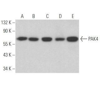
- PAK4 Antibody (B-3) is a mouse monoclonal IgG1 κ PAK4 antibody, cited in 12 publications, provided at 200 µg/ml
- specific for an epitope mapping between amino acids 167-209 within an internal region of PAK4 of human origin
- PAK4 Antibody (B-3) is recommended for detection of PAK4 of mouse, rat and human origin by WB, IP, IF, IHC(P) and ELISA
- Anti-PAK4 Antibody (B-3) is available conjugated to agarose for IP; HRP for WB, IHC(P) and ELISA; and to either phycoerythrin or FITC for IF, IHC(P) and FCM
- also available conjugated to Alexa Fluor® 488, Alexa Fluor® 546, Alexa Fluor® 594 or Alexa Fluor® 647 for WB (RGB), IF, IHC(P) and FCM, and for use with RGB fluorescent imaging systems, such as iBright™ FL1000, FluorChem™, Typhoon, Azure and other comparable systems
- also available conjugated to Alexa Fluor® 680 or Alexa Fluor® 790 for WB (NIR), IF and FCM; for use with Near-Infrared (NIR) detection systems, such as LI-COR®Odyssey®, iBright™ FL1000, FluorChem™, Typhoon, Azure and other comparable systems
- m-IgG Fc BP-HRP and m-IgG1 BP-HRP are the preferred secondary detection reagents for PAK4 Antibody (B-3) for WB and IHC(P) applications. These reagents are now offered in bundles with PAK4 Antibody (B-3) (see ordering information below).

QUICK LINKS
SEE ALSO...
PAK4 Antibody (B-3) is a mouse monoclonal IgG1 kappa light chain antibody that detects PAK4 protein of mouse, rat, and human origin by western blotting (WB), immunoprecipitation (IP), immunofluorescence (IF), immunohistochemistry, and enzyme-linked immunosorbent assay (ELISA). PAK4 Antibody (B-3) is available in both non-conjugated and various conjugated forms, including agarose, horseradish peroxidase (HRP), phycoerythrin (PE), fluorescein isothiocyanate (FITC), and multiple Alexa Fluor® conjugates. PAK4, a member of the serine/threonine kinase family, plays a crucial role in cellular signaling pathways, particularly in the regulation of cytoskeletal dynamics and cell motility. PAK4 is primarily located in the cytoplasm and interacts with GTP-bound forms of the small GTPases Rac1 and Cdc42, which are essential for actin filament organization. PAK4 interaction with these GTPases is vital for the formation of actin-enriched filopodia, structures that facilitate cell movement and communication. PAK4 is implicated in the activation of the c-Jun N-terminal kinase (JNK) pathway, which is involved in various cellular processes, including stress responses and apoptosis. PAK4 gene is located on human chromosome 19, with notably high expression in tissues such as the prostate, testis, and colon, highlighting PAK4′s potential significance in both normal physiology and disease states.
Alexa Fluor® is a trademark of Molecular Probes Inc., OR., USA
LI-COR® and Odyssey® are registered trademarks of LI-COR Biosciences
PAK4 Antibody (B-3) References:
- Molecular cloning of the gene for the human placental GTP-binding protein Gp (G25K): identification of this GTP-binding protein as the human homolog of the yeast cell-division-cycle protein CDC42. | Shinjo, K., et al. 1990. Proc Natl Acad Sci U S A. 87: 9853-7. PMID: 2124704
- Regulation of KRAS-PAK4 axis by microRNAs in cancer. | Choudhry, ZS., et al. 2014. Curr Pharm Des. 20: 5275-8. PMID: 24479809
- rac, a novel ras-related family of proteins that are botulinum toxin substrates. | Didsbury, J., et al. 1989. J Biol Chem. 264: 16378-82. PMID: 2674130
- The role of PAK4 in the immune system and its potential implication in cancer immunotherapy. | Naїja, A., et al. 2021. Cell Immunol. 367: 104408. PMID: 34246086
- Targeting PAK4 reverses cisplatin resistance in NSCLC by modulating ER stress. | Liu, S., et al. 2024. Cell Death Discov. 10: 36. PMID: 38238316
- The small GTP-binding proteins Rac1 and Cdc42 regulate the activity of the JNK/SAPK signaling pathway. | Coso, OA., et al. 1995. Cell. 81: 1137-46. PMID: 7600581
- A novel serine kinase activated by rac1/CDC42Hs-dependent autophosphorylation is related to PAK65 and STE20. | Martin, GA., et al. 1995. EMBO J. 14: 1970-8. PMID: 7744004
- Activation of stress-activated protein kinase by MEKK1 phosphorylation of its activator SEK1. | Yan, M., et al. Nature. 372: 798-800. PMID: 7997270
- A brain serine/threonine protein kinase activated by Cdc42 and Rac1. | Manser, E., et al. 1994. Nature. 367: 40-6. PMID: 8107774
- Proteins regulating Ras and its relatives. | Boguski, MS. and McCormick, F. 1993. Nature. 366: 643-54. PMID: 8259209
- A divergence in the MAP kinase regulatory network defined by MEK kinase and Raf. | Lange-Carter, CA., et al. 1993. Science. 260: 315-9. PMID: 8385802
- PAK4, a novel effector for Cdc42Hs, is implicated in the reorganization of the actin cytoskeleton and in the formation of filopodia. | Abo, A., et al. 1998. EMBO J. 17: 6527-40. PMID: 9822598
Ordering Information
| Product Name | Catalog # | UNIT | Price | Qty | FAVORITES | |
PAK4 Antibody (B-3) | sc-390507 | 200 µg/ml | $322.00 | |||
PAK4 Antibody (B-3): m-IgG Fc BP-HRP Bundle | sc-527637 | 200 µg Ab; 10 µg BP | $361.00 | |||
PAK4 Antibody (B-3): m-IgG1 BP-HRP Bundle | sc-533010 | 200 µg Ab; 20 µg BP | $361.00 | |||
PAK4 Antibody (B-3) AC | sc-390507 AC | 500 µg/ml, 25% agarose | $424.00 | |||
PAK4 Antibody (B-3) HRP | sc-390507 HRP | 200 µg/ml | $322.00 | |||
PAK4 Antibody (B-3) FITC | sc-390507 FITC | 200 µg/ml | $336.00 | |||
PAK4 Antibody (B-3) PE | sc-390507 PE | 200 µg/ml | $349.00 | |||
PAK4 Antibody (B-3) Alexa Fluor® 488 | sc-390507 AF488 | 200 µg/ml | $364.00 | |||
PAK4 Antibody (B-3) Alexa Fluor® 546 | sc-390507 AF546 | 200 µg/ml | $364.00 | |||
PAK4 Antibody (B-3) Alexa Fluor® 594 | sc-390507 AF594 | 200 µg/ml | $364.00 | |||
PAK4 Antibody (B-3) Alexa Fluor® 647 | sc-390507 AF647 | 200 µg/ml | $364.00 | |||
PAK4 Antibody (B-3) Alexa Fluor® 680 | sc-390507 AF680 | 200 µg/ml | $364.00 | |||
PAK4 Antibody (B-3) Alexa Fluor® 790 | sc-390507 AF790 | 200 µg/ml | $364.00 | |||
PAK4 (B-3) Neutralizing Peptide | sc-390507 P | 100 µg/0.5 ml | $69.00 |