")
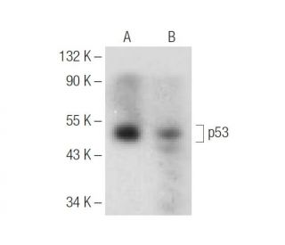
: sc-374087. Analyse par Western blot de l'expression de p53 dans des lysats de cellules entières de WR19L (A) et de souris LacZ (B).")
: sc-374087. Coloration par immunofluorescence de cellules NIH/3T3 fixées au méthanol montrant une localisation nucléaire et cytoplasmique.")
: sc-374087. Analyse par Western blot de l'expression de p53 dans des lysats de cellules entières WR19L (A), F9 (B) et T-47D (C).")
Anticorps p53 (F-8): sc-374087
- L'Anticorps p53 (F-8) est un monoclonal de souris IgG1 κ, cité dans 5 publications, fourni en 200 µg/ml
- spécifique d'un épitope situé entre les acides aminés 367-390 au niveau C-terminus de p53 d'origine mouse
- recommandé pour la détection de p53 d'origine mouse, rat et human par WB, IP, IF et ELISA
- Voir p53 (DO-1): sc-126 pour d'autres anticorps conjugués p53, dont AC, HRP, FITC, PE, Alexa Fluor® 488, 594, 647, 680 et 790.
- 1 BP-HRP">m-IgG1 BP-HRP et m-IgGκ BP-HRP sont les réactifs de détection secondaire préférés pour l'anticorps p53 (F-8) pour les applications WB. Ces réactifs sont désormais proposés en lots avec l'anticorps p53 (F-8)(voir les informations relatives à la commande ci-dessous).

ACCÈS RAPIDE AUX LIENS
VOIR ÉGALEMENT...
Anticorps p53 (F-8) IgG1 monoclonal de chaîne légère kappa de souris spécifique d'un épitope situé entre les acides aminés 367-390 à l'extrémité C-terminale de la protéine p53 d'origine murine. L'anticorps monoclonal p53 (F-8) réagit avec les espèces de souris, de rats et d'humains et est recommandé pour diverses applications telles que le western blotting, l'immunoprécipitation, l'immunofluorescence et l'enzyme-linked immunosorbent assay (essai immunoenzymatique). La protéine p53, codée par le gène TP53, est un suppresseur de tumeur essentiel qui fonctionne principalement comme un facteur de transcription régulant des processus clés, notamment la réparation de l'ADN, l'apoptose, l'arrêt du cycle cellulaire et la sénescence. Lorsque l'ADN est endommagé, p53 peut arrêter le cycle cellulaire au point de contrôle G1/S, ce qui laisse le temps aux mécanismes de réparation de corriger les dommages. Si les dommages sont irréparables, p53 peut déclencher l'apoptose, empêchant ainsi la propagation des cellules mutées. La recherche sur les anticorps p53, en particulier l'anticorps monoclonal p53 (F-8), s'est avérée inestimable pour étudier la localisation et l'expression de p53 dans les cellules. L'anticorps de souris p53 (F-8) se lie spécifiquement à p53, ce qui permet aux chercheurs d'étudier l'implication de p53 dans le développement et la progression du cancer. Étant donné que des mutations du gène TP53 sont présentes dans près de la moitié des cancers humains, la restauration ou la modulation de la fonction de p53 reste un axe essentiel de la recherche sur les thérapies anticancéreuses. Les mutations de p53, en particulier dans son domaine de liaison à l'ADN, entraînent souvent une perte de suppression de la tumeur et peuvent conférer des propriétés oncogènes qui favorisent la croissance de la tumeur. L'anticorps anti-p53 (F-8) joue un rôle essentiel dans la recherche sur le cancer, en facilitant l'identification et l'étude des mutations de p53 dans divers types de cancer. En comprenant comment les différentes mutations affectent la structure et la fonction de p53, les scientifiques sont mieux équipés pour développer des thérapies ciblées visant à réactiver p53 ou à inhiber ses régulateurs négatifs tels que MDM2.
Alexa Fluor® est une marque déposée de Molecular Probes Inc., OR., USA
LI-COR® et Odyssey® sont marques déposées de LI-COR Biosciences
Anticorps p53 (F-8) Références:
- La 9-Hydroxyellipticine modifie la conformation et les caractéristiques de liaison à l'ADN de la protéine p53 mutée. | Sugikawa, E., et al. 2001. Anticancer Res. 21: 2671-5. PMID: 11724337
- Le gène suppresseur de tumeur TP53 : implications pour la gestion et la thérapie du cancer. | Seemann, S., et al. 2004. Crit Rev Clin Lab Sci. 41: 551-83. PMID: 15603511
- Un anticorps à chaîne unique contre l'épitope commun de la protéine p53 mutante restaure l'activité de type sauvage de la protéine p53 mutante. | Orgad, S., et al. 2005. FEBS Lett. 579: 5609-15. PMID: 16213502
- Vers la compréhension de la reconnaissance moléculaire de l'albumine par le peptide agrafé activateur de p53 ATSP-7041. | Tiwari, G. and Verma, CS. 2017. J Phys Chem B. 121: 657-670. PMID: 28048940
- Formes et fonctions de p53. | Milner, J. 1994. Semin Cancer Biol. 5: 211-9. PMID: 7948949
- Gènes suppresseurs de tumeurs et chaperons moléculaires. | Lane, DP., et al. 1993. Philos Trans R Soc Lond B Biol Sci. 339: 369-72; discussion 372-3. PMID: 8493291
- L'antisens p53 provoque des changements dans la croissance et la morphologie des cellules HeLa. | Iotsova, V. and Stehelin, D. 1995. Eur J Cell Biol. 68: 122-32. PMID: 8575459
- Déterminations sérologiques et immunohistochimiques de la protéine p53 dans le diagnostic du cancer de l'endomètre : une étude comparative. | Ben-Hur, H., et al. 1997. Eur J Gynaecol Oncol. 18: 400-6. PMID: 9378162
- La fonction et la conformation de la protéine p53 de type sauvage sont influencées par des mutations dans le lymphosarcome à cellules B induit par le virus de la leucémie bovine. | Tajima, S., et al. 1998. Virology. 243: 735-46. PMID: 9540787
- La curcumine induit une apoptose dépendante de p53 dans les cellules de carcinome basocellulaire humain. | Jee, SH., et al. 1998. J Invest Dermatol. 111: 656-61. PMID: 9764849
Informations pour la commande
| Nom du produit | Ref. Catalogue | COND. | Prix HT | QTÉ | Favoris | |
Anticorps p53 (F-8) | sc-374087 | 200 µg/ml | $322.00 | |||
p53 (F-8): m-IgGκ BP-HRP Kit | sc-535381 | 200 µg Ab; 40 µg BP | $361.00 | |||
p53 (F-8): m-IgG1 BP-HRP Kit | sc-538884 | 200 µg Ab; 20 µg BP | $361.00 | |||
p53 (F-8) peptide neutralisant | sc-374087 P | 100 µg/0.5 ml | $69.00 |