
Date published: 2026-5-29

1-800-457-3801

SCBT Portrait Logo
Seach Input

p-Src Antibody (H-3): sc-166860

4.7(19)
Write a reviewAsk a question

Datasheets
  • p-Src Antibody (H-3) is a mouse monoclonal IgG2b κ, cited in 26 publications, provided at 200 µg/ml
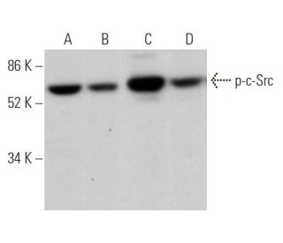
  • p-Src Antibody (H-3) is recommended for detection of Tyr 530 phosphorylated c-Src of human and rat origin, and Tyr 535 phosphorylated c-Src of mouse origin by WB, IP, IF, IHC(P) and ELISA
  • Anti-p-Src Antibody (H-3) is available conjugated to agarose for IP; HRP for WB, IHC(P) and ELISA; and to either phycoerythrin or FITC for IF, IHC(P) and FCM
  • also available conjugated to Alexa Fluor® 488, Alexa Fluor® 546, Alexa Fluor® 594 or Alexa Fluor® 647 for WB (RGB), IF, IHC(P) and FCM, and for use with RGB fluorescent imaging systems, such as iBright™ FL1000, FluorChem™, Typhoon, Azure and other comparable systems
  • also available conjugated to Alexa Fluor® 680 or Alexa Fluor® 790 for WB (NIR), IF and FCM; for use with Near-Infrared (NIR) detection systems, such as LI-COR®Odyssey®, iBright™ FL1000, FluorChem™, Typhoon, Azure and other comparable systems
  • m-IgG2b BP-HRP and m-IgGκ BP-HRP are the preferred secondary detection reagents for p-Src Antibody (H-3) for WB and IHC(P) applications. These reagents are now offered in bundles with p-Src Antibody (H-3) (see ordering information below).
Gene Editing Promo Banner

QUICK LINKS

SEE ALSO...

p-c-Src Antibody (H-3) is a mouse monoclonal IgG2b kappa light chain antibody raised against the pY530 site of human c-Src and pY535 site of mouse c-Src, with corresponding recognition of rat pY530, a critical regulatory modification of this tyrosine kinase. p-c-Src mouse monoclonal antibody (H-3) recognizes c-Src only when these specific tyrosine residues are phosphorylated and demonstrates effectiveness in Western blotting (WB), immunoprecipitation (IP), immunofluorescence (IF), immunohistochemistry on paraffin-embedded sections (IHCP), and enzyme-linked immunosorbent assay (ELISA) applications. Anti-p-c-Src monoclonal antibody (H-3) is available in non-conjugated form and multiple conjugated forms including agarose (Ag), horseradish peroxidase (HRP), phycoerythrin (PE), fluorescein isothiocyanate (FITC), and various Alexa Fluor® (AF) conjugates, offering versatility for diverse experimental needs. As a member of the Src family kinases, c-Src primarily resides in the cytoplasm but can undergo translocation to the plasma membrane in response to various stimuli. This dynamic localization enables c-Src to interact with membrane-associated proteins and participate in signaling cascades that regulate cellular responses to growth factors and other external signals. c-Src typically maintains an inactive state until specific cellular events, such as mitosis, trigger c-Src activation, highlighting c-Src′s importance in cell cycle progression and other dynamic cellular processes. Through c-Src′s broad expression pattern, particularly in neuronal tissues and platelets, c-Src plays crucial roles in neuronal differentiation and mature neuronal function maintenance. Through these diverse cellular functions, p-c-Src (H-3) antibody serves as an essential tool for researchers investigating signaling pathways that control cell proliferation, differentiation, and motility.

For Research Use Only. Not Intended for Diagnostic or Therapeutic Use.

Alexa Fluor® is a trademark of Molecular Probes Inc., OR., USA

LI-COR® and Odyssey® are registered trademarks of LI-COR Biosciences

p-Src Antibody (H-3) References:

  1. Herpesvirus saimiri protein StpB associates with cellular Src.  |  Hör, S., et al. 2001. J Gen Virol. 82: 339-344. PMID: 11161272
  2. Regulation of the oncogenic activity of the cellular src protein requires the correct spacing between the kinase domain and the C-terminal phosphorylated tyrosine (Tyr-527).  |  Cobb, BS., et al. 1991. Mol Cell Biol. 11: 5832-8. PMID: 1719372
  3. Phosphorylated c-Src in the nucleus is associated with improved patient outcome in ER-positive breast cancer.  |  Campbell, EJ., et al. 2008. Br J Cancer. 99: 1769-74. PMID: 19018258
  4. Phosphorylated c-Src is a novel predictor for recurrence in cervical squamous cell cancer patients.  |  Hou, T., et al. 2013. Int J Clin Exp Pathol. 6: 1121-7. PMID: 23696930
  5. The cellular src gene product regulates junctional cell-to-cell communication.  |  Azarnia, R., et al. 1988. Science. 239: 398-401. PMID: 2447651
  6. Sphingosine kinase 2 prevents the nuclear translocation of sphingosine 1-phosphate receptor-2 and tyrosine 416 phosphorylated c-Src and increases estrogen receptor negative MDA-MB-231 breast cancer cell growth: The role of sphingosine 1-phosphate receptor-4.  |  Ohotski, J., et al. 2014. Cell Signal. 26: 1040-7. PMID: 24486401
  7. Extracellular Vesicle Activation of Latent HIV-1 Is Driven by EV-Associated c-Src and Cellular SRC-1 via the PI3K/AKT/mTOR Pathway.  |  Barclay, RA., et al. 2020. Viruses. 12: PMID: 32575590
  8. Phospholipase C-gamma 1 associates with viral and cellular src kinases.  |  Nakanishi, O., et al. 1993. J Biol Chem. 268: 10754-9. PMID: 8388373
  9. Association of tyrosine-phosphorylated c-Src with the cytoskeleton of hypertrophying myocardium.  |  Kuppuswamy, D., et al. 1997. J Biol Chem. 272: 4500-8. PMID: 9020175
  10. Genetic variation of herpesvirus saimiri subgroup A transforming protein and its association with cellular src.  |  Lee, H., et al. 1997. J Virol. 71: 3817-25. PMID: 9094657

Ordering Information

Product NameCatalog #UNITPriceQtyFAVORITES

p-Src Antibody (H-3)

sc-166860
200 µg/ml
$322.00

p-Src Antibody (H-3): m-IgGκ BP-HRP Bundle

sc-521777
200 µg Ab, 40 µg BP
$361.00

p-Src Antibody (H-3): m-IgG2b BP-HRP Bundle

sc-548965
200 µg Ab; 10 µg BP
$361.00

p-Src Antibody (H-3) AC

sc-166860 AC
500 µg/ml, 25% agarose
$424.00

p-Src Antibody (H-3) HRP

sc-166860 HRP
200 µg/ml
$322.00

p-Src Antibody (H-3) FITC

sc-166860 FITC
200 µg/ml
$336.00

p-Src Antibody (H-3) PE

sc-166860 PE
200 µg/ml
$349.00

p-Src Antibody (H-3) Alexa Fluor® 488

sc-166860 AF488
200 µg/ml
$364.00

p-Src Antibody (H-3) Alexa Fluor® 546

sc-166860 AF546
200 µg/ml
$364.00

p-Src Antibody (H-3) Alexa Fluor® 594

sc-166860 AF594
200 µg/ml
$364.00

p-Src Antibody (H-3) Alexa Fluor® 647

sc-166860 AF647
200 µg/ml
$364.00

p-Src Antibody (H-3) Alexa Fluor® 680

sc-166860 AF680
200 µg/ml
$364.00

p-Src Antibody (H-3) Alexa Fluor® 790

sc-166860 AF790
200 µg/ml
$364.00

p-Src (H-3) Neutralizing Peptide

sc-166860 P
100 µg/0.5 ml
$69.00

What application is the blocking peptide sc-166860 P appropriate for?

Asked by: jenniferc
Thank you for your question. The blocking peptide is intended for use as a negative control, by pre-adsorbing the mouse monoclonal antibody against the antigen. For full protocol details, please contact our Technical Services Department or view our online protocol here: https://www.scbt.com/scbt/resources/protocols/peptide-neutralization
Answered by: Technical Support
Date published: 2017-02-24
  • y_2026, m_5, d_28, h_14CST
  • bvseo_bulk, prod_bvqa, vn_bulk_3.0.46
  • cp_1, bvpage1
  • co_hasquestionsanswers, tq_1
  • loc_en_US, sid_166860, prod, sort_[SortEntry(order=LAST_APPROVED_ANSWER_SUBMISSION_TIME, direction=DESCENDING)]
  • clientName_scbt
  • bvseo_sdk, java_sdk, bvseo-4.0.0
  • CLOUD, getContent, 100ms
  • QUESTIONS, PRODUCT
Rated 5 out of 5 by from Worked well in WBThis antibody worked well in WB using human macrophages
Date published: 2025-06-06
Rated 5 out of 5 by from Worked for human tissue samples WBWorked well for human colon cancer cell lysates using WB at 1:200. Had to expose for longer period of time though.
Date published: 2018-03-22
Rated 4 out of 5 by from p-c-Src Antibody(H-3)H-3 is the ideal ab to detect p-c-Src of my sample, but it needs high concentration to start with.
Date published: 2018-03-07
Rated 5 out of 5 by from best phospho srcuse in a experiment of WB. it works very well for me
Date published: 2018-02-21
Rated 3 out of 5 by from seems not good for liver sample testI try to test healthy liver samples, but seems only a very specific band with about 38Kd, but no the target band.
Date published: 2017-12-04
Rated 5 out of 5 by from Good antibodyGives me strong and specific bands. I would recommend it.
Date published: 2017-09-23
Rated 5 out of 5 by from works great on western blot and IHCWe have used this antibody on human skin tissue. It works great on both western blot and IHC.
Date published: 2017-09-20
Rated 5 out of 5 by from sharp bandsThis antibody was tested on human aortic endothelial cells post various treatments. It gives specific band with no background. A non-specific band around 40 kDa was observed.
Date published: 2017-09-14
  • y_2026, m_5, d_28, h_14
  • bvseo_bulk, prod_bvrr, vn_bulk_3.0.46
  • cp_1, bvpage1
  • co_hasreviews, tv_0, tr_19
  • loc_en_US, sid_166860, prod, sort_[SortEntry(order=SUBMISSION_TIME, direction=DESCENDING)]
  • clientName_scbt
  • bvseo_sdk, java_sdk, bvseo-4.0.0
  • CLOUD, getReviews, 10ms
  • REVIEWS, PRODUCT
p-Src Antibody (H-3) is rated 4.7 out of 5 by 19.
  • y_2026, m_5, d_28, h_14
  • bvseo_bulk, prod_bvrr, vn_bulk_3.0.46
  • cp_1, bvpage1
  • co_hasreviews, tv_0, tr_19
  • loc_en_US, sid_166860, prod, sort_[SortEntry(order=SUBMISSION_TIME, direction=DESCENDING)]
  • clientName_scbt
  • bvseo_sdk, java_sdk, bvseo-4.0.0
  • CLOUD, getAggregateRating, 98ms
  • REVIEWS, PRODUCT
