")
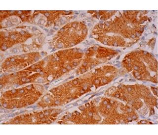
: sc-377375. Immunoperoxidase staining of formalin fixed, paraffin-embedded human upper stomach tissue showing cytoplasmic staining of glandular cells.")
: sc-377375. Western blot analysis of MIA expression in non-transfected: sc-117752 (A) and mouse MIA transfected: sc-127154 (B) 293T whole cell lysates.")
MIA Antibody (C-10): sc-377375
- MIA Antibody (C-10) is a mouse monoclonal IgG2b κ MIA antibody, cited in 3 publications, provided at 200 µg/ml
- raised against amino acids 62-131 mapping at the C-terminus of MIA of human origin
- MIA Antibody (C-10) is recommended for detection of precursor and mature forms of MIA of mouse, rat and human origin by WB, IP, IF, IHC(P) and ELISA
- Anti-MIA Antibody (C-10) is available conjugated to agarose for IP; HRP for WB, IHC(P) and ELISA; and to either phycoerythrin or FITC for IF, IHC(P) and FCM
- also available conjugated to Alexa Fluor® 488, Alexa Fluor® 546, Alexa Fluor® 594 or Alexa Fluor® 647 for WB (RGB), IF, IHC(P) and FCM, and for use with RGB fluorescent imaging systems, such as iBright™ FL1000, FluorChem™, Typhoon, Azure and other comparable systems
- also available conjugated to Alexa Fluor® 680 or Alexa Fluor® 790 for WB (NIR), IF and FCM; for use with Near-Infrared (NIR) detection systems, such as LI-COR®Odyssey®, iBright™ FL1000, FluorChem™, Typhoon, Azure and other comparable systems
- m-IgGκ BP-HRP is the preferred secondary detection reagent for MIA Antibody (C-10) for WB and IHC(P) applications. This reagent is now offered in a bundle with MIA Antibody (C-10) (see ordering information below). For additional m-IgGκ BP conjugates see our complete list of Mouse IgG Binding Proteins.

QUICK LINKS
MIA Antibody (C-10) is a mouse monoclonal IgG2b kappa light chain antibody that detects MIA protein of mouse, rat, and human origin by western blotting (WB), immunoprecipitation (IP), immunofluorescence (IF), immunohistochemistry, and enzyme-linked immunosorbent assay (ELISA). anti-MIA antibody (C-10) is available in both non-conjugated and various conjugated forms, including agarose, horseradish peroxidase (HRP), phycoerythrin (PE), fluorescein isothiocyanate (FITC), and multiple Alexa Fluor® conjugates. MIA protein, also known as cartilage-derived retinoic acid-sensitive protein, plays a crucial role in tumorigenesis, particularly in malignant melanomas, by influencing cell proliferation and interaction with the extracellular matrix through complex signaling pathways. MIA is secreted by chondrocytes and malignant melanoma cells, and is initially synthesized as a 131-amino acid precursor that is subsequently processed into a mature 107-amino acid form following secretion signal cleavage. MIA protein is not only essential during chondrogenesis and in maintaining normal phenotype of mature chondrocytes, but is also implicated in various carcinomas, including those of colon, ovary, kidney, and head/neck, making MIA a potential biomarker for monitoring melanomic activity. MIA′s ability to modulate tumor microenvironment highlights its significance in cancer biology, providing insights into therapeutic targets and diagnostic markers for melanoma and other malignancies.
Alexa Fluor® is a trademark of Molecular Probes Inc., OR., USA
LI-COR® and Odyssey® are registered trademarks of LI-COR Biosciences
MIA Antibody (C-10) References:
- Expression of melanoma inhibitory activity in melanoma and nonmelanoma tissue specimens. | Perez, RP., et al. 2000. Hum Pathol. 31: 1381-8. PMID: 11112213
- The extracellular human melanoma inhibitory activity (MIA) protein adopts an SH3 domain-like fold. | Stoll, R., et al. 2001. EMBO J. 20: 340-9. PMID: 11157741
- Structure of melanoma inhibitory activity protein, a member of a recently identified family of secreted proteins. | Lougheed, JC., et al. 2001. Proc Natl Acad Sci U S A. 98: 5515-20. PMID: 11331761
- Cloning of a novel malignant melanoma-derived growth-regulatory protein, MIA. | Blesch, A., et al. 1994. Cancer Res. 54: 5695-701. PMID: 7923218
- Mouse CD-RAP/MIA gene: structure, chromosomal localization, and expression in cartilage and chondrosarcoma. | Bosserhoff, AK., et al. 1997. Dev Dyn. 208: 516-25. PMID: 9097023
Ordering Information
| Product Name | Catalog # | UNIT | Price | Qty | FAVORITES | |
MIA Antibody (C-10) | sc-377375 | 200 µg/ml | $322.00 | |||
MIA Antibody (C-10): m-IgGκ BP-HRP Bundle | sc-523302 | 200 µg Ab, 40 µg BP | $361.00 | |||
MIA Antibody (C-10) AC | sc-377375 AC | 500 µg/ml, 25% agarose | $424.00 | |||
MIA Antibody (C-10) HRP | sc-377375 HRP | 200 µg/ml | $322.00 | |||
MIA Antibody (C-10) FITC | sc-377375 FITC | 200 µg/ml | $336.00 | |||
MIA Antibody (C-10) PE | sc-377375 PE | 200 µg/ml | $349.00 | |||
MIA Antibody (C-10) Alexa Fluor® 488 | sc-377375 AF488 | 200 µg/ml | $364.00 | |||
MIA Antibody (C-10) Alexa Fluor® 546 | sc-377375 AF546 | 200 µg/ml | $364.00 | |||
MIA Antibody (C-10) Alexa Fluor® 594 | sc-377375 AF594 | 200 µg/ml | $364.00 | |||
MIA Antibody (C-10) Alexa Fluor® 647 | sc-377375 AF647 | 200 µg/ml | $364.00 | |||
MIA Antibody (C-10) Alexa Fluor® 680 | sc-377375 AF680 | 200 µg/ml | $364.00 | |||
MIA Antibody (C-10) Alexa Fluor® 790 | sc-377375 AF790 | 200 µg/ml | $364.00 |