")
JAM-A Anticuerpo (J3F.1): sc-53622
- JAM-A Anticuerpo (J3F.1) es un monoclonal de ratón IgG1 κ, ver las 5 publicaciones, proporcionado como 200 µg/ml
- planteada frente a la proteína de fusión JAM recombinante de origen human.
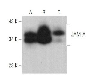
- recomendado para detectar JAM-A de human origen, mediante WB, IP, IF, IHC(P) y FCM
- disponible conjugado tanto a ficoeritrina como a FITC para IF, IHC(P) y FCM
- Ver JAM-A (J10.4): sc-53623 para JAM-A anticuerpos conjugados, incluyendo AC, HRP, FITC, PE, Alexa Fluor® 488, 594, 647, 680 y 790.
- m-IgG Fc BP-HRP y m-IgG1 BP-HRP son los reactivos de detección secundarios preferidos para JAM-A Anticuerpo (J3F.1) for WB and IHC(P) applications. Estos reactivos se ofrecen ahora en paquetes con JAM-A Anticuerpo (J3F.1)(véase la información de pedido más abajo).

La molécula de adhesión juncional (JAM) es un miembro de la superfamilia de inmunoglobulinas expresada en uniones estrechas de células epiteliales y células endoteliales. Está implicada en la migración transendotelial de leucocitos. JAM se expresa de forma constitutiva en monocitos circulantes, neutrófilos, subconjuntos de linfocitos y plaquetas. La familia JAM consta de JAM-A, JAM-B y JAM-C, designados alternativamente como JAM-1, JAM-2 y JAM-3, respectivamente. JAM-A se localiza con F-Actina en los contactos célula-célula y en los pliegues de la membrana. Está involucrada en la adhesión célula a célula a través de interacciones homofílicas y desempeña un papel en la organización de uniones estrechas y la modulación de la extravasación de leucocitos. JAM-B interactúa con subconjuntos discretos de linfocitos periféricos, lo que sugiere que puede desempeñar un papel en el tráfico de linfocitos. Las proteínas JAM-B y JAM-C son socios de unión; JAM-C puede ser un receptor funcional de JAM-B. Específicamente, JAM-B se adhiere a las células T a través de interacciones heterotípicas con JAM-C. La interacción JAM-B/JAM-C puede desempeñar un papel en la inflamación celular de células T, NK y dendríticas.
Información sobre pedidos
| Nombre del producto | Número de catálogo | UNIDAD | Precio | CANTIDAD | Favoritos | |
JAM-A Anticuerpo (J3F.1) | sc-53622 | 200 µg/ml | $322.00 | |||
Paquete de JAM-A (J3F.1): m-IgG Fc BP-HRP | sc-538999 | 200 µg Ab; 10 µg BP | $361.00 | |||
Paquete de JAM-A (J3F.1): m-IgG1 BP-HRP | sc-541200 | 200 µg Ab; 20 µg BP | $361.00 | |||
JAM-A Anticuerpo (J3F.1) FITC | sc-53622 FITC | 200 µg/ml | $336.00 | |||
JAM-A Anticuerpo (J3F.1) PE | sc-53622 PE | 200 µg/ml | $349.00 |