")
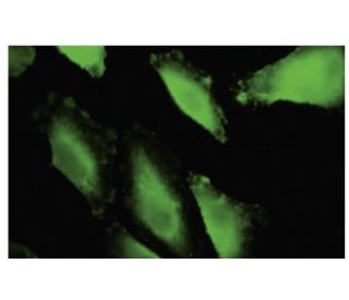
: sc-101124. Immunofluorescence staining of paraformaldehyde-fixed Hep G2 cells showing cytoplasmic localization.")
: sc-101124. Western blot analysis of IBRDC1 expression in Hep G2 whole cell lysate.")
IBRDC1 Antibody (36-A2): sc-101124
- IBRDC1 Antibody (36-A2) is a mouse monoclonal IgG2a κ IBRDC1 antibody provided at 50 µg/0.5 ml
- raised against recombinant IBRDC1 of human origin
- recommended for detection of IBRDC1 of mouse, rat and human origin by WB, IP, IF and ELISA
- At present, we have not yet completed the identification of the preferred secondary detection reagent(s) for IBRDC1 Antibody (36-A2). This work is in progress.

IBRDC1 Antibody (36-A2) is a mouse monoclonal IgG2a kappa light chain antibody that detects IBRDC1 protein of mouse, rat, and human origin by western blotting (WB), immunoprecipitation (IP), immunofluorescence (IF), and enzyme-linked immunosorbent assay (ELISA). Anti-IBRDC1 antibody (36-A2) is available as non-conjugated format. IBRDC1, also known as RNF217, is a 275 amino acid single-pass membrane protein characterized by two IBR (in between ring fingers)-type zinc finger motifs, which are crucial for function as an E3 ubiquitin ligase. IBRDC1 plays a significant role in ubiquitin-proteasome pathway, a vital cellular mechanism responsible for regulating protein degradation and turnover. IBRDC1′s ability to mediate protein ubiquitinylation is essential for maintaining cellular homeostasis, influencing processes such as cell cycle progression, signal transduction, and stress response. Through targeted protein degradation, IBRDC1 helps control levels of key regulatory proteins, impacting cellular functions and overall organismal health. IBRDC1′s versatility in modulating protein interactions and stability underscores importance in cellular regulation and potential implications in disease states where these processes are disrupted.
Alexa Fluor® is a trademark of Molecular Probes Inc., OR., USA
LI-COR® and Odyssey® are registered trademarks of LI-COR Biosciences
IBRDC1 Antibody (36-A2) References:
- Features of the parkin/ariadne-like ubiquitin ligase, HHARI, that regulate its interaction with the ubiquitin-conjugating enzyme, Ubch7. | Ardley, HC., et al. 2001. J Biol Chem. 276: 19640-7. PMID: 11278816
- Parkin and relatives: the RBR family of ubiquitin ligases. | Marín, I., et al. 2004. Physiol Genomics. 17: 253-63. PMID: 15152079
- Comparative genomics and protein domain graph analyses link ubiquitination and RNA metabolism. | Lucas, JI., et al. 2006. J Mol Biol. 357: 9-17. PMID: 16426638
- UVPAR: fast detection of functional shifts in duplicate genes. | Arnau, V., et al. 2006. BMC Bioinformatics. 7: 174. PMID: 16569227
- Structure of the Parkin in-between-ring domain provides insights for E3-ligase dysfunction in autosomal recessive Parkinson's disease. | Beasley, SA., et al. 2007. Proc Natl Acad Sci U S A. 104: 3095-100. PMID: 17360614
- The ring between ring fingers (RBR) protein family. | Eisenhaber, B., et al. 2007. Genome Biol. 8: 209. PMID: 17367545
Ordering Information
| Product Name | Catalog # | UNIT | Price | Qty | FAVORITES | |
IBRDC1 Antibody (36-A2) | sc-101124 | 50 µg/0.5 ml | $322.00 |