")
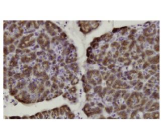
: sc-100720. Immunoperoxidasefärbung von formalinfixiertem, paraffineingebettetem menschlichem Pankreasgewebe mit zytoplasmatischer Lokalisierung.")
Herc3 Antikörper (37.2): sc-100720
- Herc3 Antikörper 37.2 ist ein Maus monoklonales IgG1 κ Herc3 Antikörper, verwendet in 1 wissenschaftlichen Veröffentlichungen, in einer Menge von 100 µg/ml
- gezogen gegen rekombinantes Herc3 aus der Spezies human
- Empfohlen für die Detektion von Herc3 aus der Spezies human per IF, IHC(P) und ELISA
- Aktuell testen wir noch unsere Sekundärantikörper um das beste Bindeprotein für diesen Primärantikörper Herc3 (37.2) zu finden. Kontaktieren Sie uns bitte, wenn Sie Fragen hierzu haben sollten.

Direktverknüpfungen
Der Herc3-Antikörper (37.2) ist ein monoklonaler Maus IgG1 κ Herc3-Antikörper (auch als Herc3-Antikörper bezeichnet), der das Herc3-Protein menschlichen Ursprungs mittels IF, IHC (P) und ELISA detektiert. Der Herc3-Antikörper (37.2) ist als nicht konjugierter Anti-Herc3-Antikörper erhältlich. Herc3 (HECT-Domäne und RCC1-ähnliche Domänen-enthaltendes Protein 3) ist ein 1050 Aminosäure-Protein, das sowohl im Cytoplasma als auch in vesikulären Strukturen lokalisiert ist. Herc3 ist an Proteinabbauwege beteiligt und fungiert als E3-Ubiquitin-Ligase, die eine Ubiquitin-Rest von einer E2-Ubiquitin-Konjugations-Enzym akzeptiert und diesen Rest sofort an ein Protein überträgt, das zur Degradation bestimmt ist. Herc3 enthält, ähnlich wie andere Mitglieder der Herc-Familie, eine HECT (E6AP-Typ E3-Ubiquitin-Protein-Ligase)-Domäne und sieben RCC1-Wiederholungen, durch die seine E3-Ubiquitin-Ligase-Aktivität übertragen wird. Nach der Ubiquitination wird Herc3 für die Proteasom-Degradation bestimmt.
Alexa Fluor® ist ein Markenzeichen von Molecular Probes Inc., OR., USA
LI-COR® und Odyssey® sind Markenzeichen von LI-COR Biosciences
Herc3 Antikörper (37.2) Literaturhinweise:
- Zuordnung des menschlichen Gens P532 (HERC1) zum Chromosom 15q22 durch Fluoreszenz-in-situ-Hybridisierung. | Cruz, C., et al. 1999. Cytogenet Cell Genet. 86: 68-9. PMID: 10516438
- Das menschliche HERC3-Gen ist durch Fluoreszenz-in-situ-Hybridisierung auf Chromosom 4q21 kartiert. | Cruz, C., et al. 1999. Cytogenet Cell Genet. 87: 263-4. PMID: 10702688
- Die menschliche HERC-Familie von Ubiquitin-Ligasen: neue Mitglieder, genomische Organisation, Expressionsprofilierung und evolutionäre Aspekte. | Hochrainer, K., et al. 2005. Genomics. 85: 153-64. PMID: 15676274
- Vorhersage der kodierenden Sequenzen von nicht identifizierten menschlichen Genen. I. Die kodierenden Sequenzen von 40 neuen Genen (KIAA0001-KIAA0040), abgeleitet durch Analyse von zufällig ausgewählten cDNA-Klonen aus der menschlichen unreifen myeloischen Zelllinie KG-1. | Nomura, N., et al. 1994. DNA Res. 1: 27-35. PMID: 7584026
Bestellinformation
| Produkt | Katalog # | EINHEIT | Preis | ANZAHL | Favoriten | |
Herc3 Antikörper (37.2) | sc-100720 | 100 µg/ml | $339.00 |