")
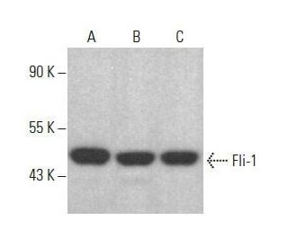
Fli-1 Antibody (F-12): sc-365294
- Fli-1 Antibody (F-12) is a mouse monoclonal IgM κ Fli-1 antibody, cited in 4 publications, provided at 200 µg/ml
- specific for an epitope mapping between amino acids 427-452 at the C-terminus of Fli-1 of mouse origin
- recommended for detection of Fli-1 of mouse, rat and human origin by WB, IP, IF and ELISA
- TransCruz reagent for ChIP application (sc-365294 X, 200 µg/0.1 ml)
- At present, we have not yet completed the identification of the preferred secondary detection reagent(s) for Fli-1 Antibody (F-12). This work is in progress.

QUICK LINKS
Fli-1 Antibody (F-12) is a mouse monoclonal IgM antibody that detects Fli-1 protein of mouse, rat, and human origin by western blotting (WB), immunoprecipitation (IP), immunofluorescence (IF), and enzyme-linked immunosorbent assay (ELISA). Anti-Fli-1 antibody (F-12) is available as the non-conjugated form. Fli-1 plays a crucial role in regulating gene expression during hematopoiesis and is involved in the development of various tissues, including the cardiovascular system. Fli-1 acts as a transcription factor that binds to specific DNA sequences, thereby influencing the expression of genes necessary for cell differentiation and proliferation. Dysregulation of Fli-1 has been implicated in several diseases, including certain types of cancer, making Fli-1 a significant target for research and therapeutic interventions. Fli-1 is part of the Ets family of transcription factors, which are characterized by a conserved DNA-binding domain that allows interaction with specific motifs in the promoter regions of target genes. Understanding Fli-1′s role in cellular processes not only sheds light on normal developmental biology but also provides insights into pathological conditions where Fli-1 function may be altered.
Alexa Fluor® is a trademark of Molecular Probes Inc., OR., USA
LI-COR® and Odyssey® are registered trademarks of LI-COR Biosciences
Fli-1 Antibody (F-12) References:
- Molecular cloning and characterization of PEA3, a new member of the Ets oncogene family that is differentially expressed in mouse embryonic cells. | Xin, JH., et al. 1992. Genes Dev. 6: 481-96. PMID: 1547944
- Ewing's sarcoma fusion protein, EWS/Fli-1 and Fli-1 protein induce PLD2 but not PLD1 gene expression by binding to an ETS domain of 5' promoter. | Kikuchi, R., et al. 2007. Oncogene. 26: 1802-10. PMID: 16964281
- Caveolin-1 (CAV1) is a target of EWS/FLI-1 and a key determinant of the oncogenic phenotype and tumorigenicity of Ewing's sarcoma cells. | Tirado, OM., et al. 2006. Cancer Res. 66: 9937-47. PMID: 17047056
- The Drosophila 74EF early puff contains E74, a complex ecdysone-inducible gene that encodes two ets-related proteins. | Burtis, KC., et al. 1990. Cell. 61: 85-99. PMID: 2107982
- elk, tissue-specific ets-related genes on chromosomes X and 14 near translocation breakpoints. | Rao, VN., et al. 1989. Science. 244: 66-70. PMID: 2539641
- Fli-1 overexpression in erythroleukemic cells promotes erythroid de-differentiation while Spi-1/PU.1 exerts the opposite effect. | Vecchiarelli-Federico, LM., et al. 2017. Int J Oncol. 51: 456-466. PMID: 28586009
- Fli-1 Governs Pericyte Dysfunction in a Murine Model of Sepsis. | Li, P., et al. 2018. J Infect Dis. 218: 1995-2005. PMID: 30053030
- Identification and preferential expression in thymic and bursal lymphocytes of a c-ets oncogene-encoded Mr 54,000 cytoplasmic protein. | Ghysdael, J., et al. 1986. Proc Natl Acad Sci U S A. 83: 1714-8. PMID: 3006066
- erg, a human ets-related gene on chromosome 21: alternative splicing, polyadenylation, and translation. | Rao, VN., et al. 1987. Science. 237: 635-9. PMID: 3299708
- Identification of Mir-182-3p/FLI-1 Axis as a Key Signaling in Immune Response in Cervical Cancer: A Comprehensive Bioinformatic Analysis. | Salmerón-Bárcenas, EG., et al. 2023. Int J Mol Sci. 24: PMID: 37047006
- The Ets1 transcription factor is widely expressed during murine embryo development and is associated with mesodermal cells involved in morphogenetic processes such as organ formation. | Kola, I., et al. 1993. Proc Natl Acad Sci U S A. 90: 7588-92. PMID: 7689222
- Effect of PU.1 phosphorylation on interaction with NF-EM5 and transcriptional activation. | Pongubala, JM., et al. 1993. Science. 259: 1622-5. PMID: 8456286
Ordering Information
| Product Name | Catalog # | UNIT | Price | Qty | FAVORITES | |
Fli-1 Antibody (F-12) | sc-365294 | 200 µg/ml | $322.00 | |||
Fli-1 (F-12) Neutralizing Peptide | sc-365294 P | 100 µg/0.5 ml | $69.00 | |||
Fli-1 Antibody (F-12) X | sc-365294 X | 200 µg/0.1 ml | $322.00 |