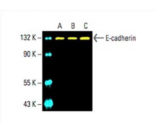
Date published: 2025-9-5

021-6093-6350

SCBT Portrait Logo
Seach Input

E-cadherin 抗体 (G-10): sc-8426

3.9(73)
寫評論提問

说明书
  • E-cadherin 抗体 G-10 是小鼠单克隆 IgG1 κ,E-cadherin抗体, 在629篇文献中引用,规格为200 µg/ml
  • 针对氨基酸600-707的抗原,在human起源的E-cadherin的细胞外域内进行定位
  • E-cadherin 抗体 (G-10) 推荐用于 WB, IP, IF, IHC(P) 和 ELISA,检测mouse, rat 和human 来源的 E-cadherin
  • 抗E-cadherin抗体(G-10)可与琼脂糖结合用于IP;与HRP结合用于WB、IHC(P)和ELISA;与藻红蛋白FITC结合用于IF、IHC(P)和FCM
  • 还可偶联Alexa Fluor® 488, Alexa Fluor® 546, Alexa Fluor® 594Alexa Fluor® 647,用于WB (RGB), IF, IHC(P) 和 FCM, 以及用于RGB荧光成像系统,例如iBright™ FL1000, FluorChem™, Typhoon, Azure和其他类似的系统
  • 还可偶联Alexa Fluor® 680Alexa Fluor® 790, 用于WB (NIR), IF 和 FCM; 以及用于近红外(NIR)检测系统,如LI-COR®/Odyssey®, iBright™ FL1000, FluorChem™, Typhoon, Azure和类似系统
  • 关于如何获取E-cadherin (G-10): sc-8426免费10 µg小样,联系我们技术服务部门 (或者您当地的代理商)了解详情。
  • m-IgG Fc BP-HRP 是 E-cadherin 抗体 (G-10) 在 WB and IHC(P) 应用中的首选二抗检测试剂。该试剂现与E-cadherin 抗体 (G-10) 组合成套装提供。 (请参阅下面的订购信息)。

快捷链接

関連項目

E-cadherin抗体(G-10)是一种小鼠单克隆IgG1 kappa轻链抗体,可通过蛋白质印迹(WB)、免疫沉淀(IP)、免疫荧光(IF)、免疫组织化学和酶联免疫吸附测定(ELISA)检测小鼠、大鼠和人类来源的E-cadherin蛋白。抗E-钙黏蛋白抗体(G-10)有非结合型和多种结合型,包括琼脂糖、辣根过氧化物酶(HRP)、藻红蛋白(PE)、异硫氰酸荧光素(FITC)和多种Alexa Fluor®结合物。E-钙粘蛋白(E-cadherin)也称为CD324或CDH1,在细胞粘附中起着至关重要的作用,而细胞粘附对于维持组织完整性和促进胚胎发育过程中的形态发生过程至关重要。E-钙黏蛋白主要位于细胞膜上,与相邻细胞形成粘附连接,从而促进组织结构稳定。E-钙黏蛋白在这些连接处的存在对于调节细胞信号通路和维持上皮细胞极性至关重要。E-钙黏蛋白功能的破坏与多种病理状况有关,包括癌症转移,其中细胞粘附力的丧失会导致细胞运动和侵袭性增强。E-cadherin与细胞质蛋白(如β-catenin)的相互作用进一步凸显了E-cadherin在细胞通讯和信号传导中的重要性,这使得E-cadherin单克隆抗体(G-10)成为研究这些关键生物学过程的宝贵工具。

仅限研究使用。不适用于诊断和治疗用途。

Alexa Fluor® 是Molecular Probes Inc., OR., USA的商标

LI-COR®和 Odyssey® 是LI-COR Biosciences的注册商标

E-cadherin 抗体 (G-10) 参考文献:

  1. 在高级别前列腺癌中,细胞粘附分子 E-cadherin 的表达减少或消失。  |  Umbas, R., et al. 1992. Cancer Res. 52: 5104-9. PMID: 1516067
  2. 小鼠 P-cadherin基因的基因组结构和染色体图谱。  |  Hatta, M., et al. 1991. Nucleic Acids Res. 19: 4437-41. PMID: 1886768
  3. [乳腺癌手术治疗]。  |  Hirsch, HA. 1978. Gynakol Rundsch. 18: 132-41. PMID: 207622
  4. 粘附蛋白:控制动物形态发生的细胞-细胞粘附分子。  |  Takeichi, M. 1988. Development. 102: 639-55. PMID: 3048970
  5. 脱丝体粘附素:另一个不断壮大的多基因粘附分子家族。  |  Koch, PJ. and Franke, WW. 1994. Curr Opin Cell Biol. 6: 682-7. PMID: 7833048
  6. 钙粘蛋白和钙凝蛋白:胚胎发育过程中的相互作用和功能。  |  Ranscht, B. 1994. Curr Opin Cell Biol. 6: 740-6. PMID: 7833053
  7. 克隆五种人类粘连蛋白明确了粘连蛋白胞外结构域的特征,并进一步证明了两种结构不同的粘连蛋白类型。  |  Tanihara, H., et al. 1994. Cell Adhes Commun. 2: 15-26. PMID: 7982033
  8. 人类内皮细胞的细胞-细胞连接中的粘附蛋白和 PECAM-1 之间的空间和时间关系。  |  Ayalon, O., et al. 1994. J Cell Biol. 126: 247-58. PMID: 8027182
  9. 粘连蛋白/卡丁蛋白复合物形成动力学:新型蛋白质相互作用和复合物组装途径。  |  Hinck, L., et al. 1994. J Cell Biol. 125: 1327-40. PMID: 8207061

订购信息

产品名称产品编号规格价格数量收藏夹

E-cadherin 抗体 (G-10)

sc-8426
200 µg/ml
$316.00

E-cadherin (G-10): m-IgG Fc BP-HRP 套装

sc-525454
200 µg Ab; 10 µg BP
$354.00

E-cadherin 抗体 (G-10) AC

sc-8426 AC
500 µg/ml, 25% agarose
$416.00

E-cadherin 抗体 (G-10) HRP

sc-8426 HRP
200 µg/ml
$316.00

E-cadherin 抗体 (G-10) FITC

sc-8426 FITC
200 µg/ml
$330.00

E-cadherin 抗体 (G-10) PE

sc-8426 PE
200 µg/ml
$343.00

E-cadherin 抗体 (G-10) Alexa Fluor® 488

sc-8426 AF488
200 µg/ml
$357.00

E-cadherin 抗体 (G-10) Alexa Fluor® 546

sc-8426 AF546
200 µg/ml
$357.00

E-cadherin 抗体 (G-10) Alexa Fluor® 594

sc-8426 AF594
200 µg/ml
$357.00

E-cadherin 抗体 (G-10) Alexa Fluor® 647

sc-8426 AF647
200 µg/ml
$357.00

E-cadherin 抗体 (G-10) Alexa Fluor® 680

sc-8426 AF680
200 µg/ml
$357.00

E-cadherin 抗体 (G-10) Alexa Fluor® 790

sc-8426 AF790
200 µg/ml
$357.00

Can I request estimate sheet ?

Asked by: Jkimlab
Thank you for your question. Please contact technical service scbt@scbt.com
Answered by: BlakeJ
Date published: 2023-07-11

can you recommend another positive tissue besides tonsil as control; please

Asked by: Lucytch
Thank you for your question. E-Cadherin is highly expressed in a number of tissues including endocrine tissues, lung, gastrointestinal tract, liver, and pancreas.
Answered by: Tech Support Europe
Date published: 2020-11-19

I'd like to use sc-8426 conjugated with Alexa Fluor488 for flow cytometry application. What isotype control antibody do you recommend to use?

Asked by: jet12
Thank you for your question. The appropriate isotype control for this antibody is normal mouse IgG1 Alexa Fluor® 488 sc-3890.
Answered by: Tech Support Europe
Date published: 2020-09-22

Hi, I used this product for IP experiments. Although I have several trials(e.g Protein G IP kit, agarose G magnetic beads and agorose G beads) I couldn't get any band for IP sample.It has H1419 lot no. Have you any recommendations(protocols, buffers etc)?

Asked by: Billgee
Thank you for your question. Please contact our Technical Service Department or your local distributor for assistance in troubleshooting this antibody. We are happy to help! Here is a link to the recommended CoA. https://www.scbt.com/resources/protocols/immunoprecipitation-western-blots
Answered by: Tech Service
Date published: 2019-12-06

Hi, ı want to learn if you have E-cadherin conjugated to agarose beads(for IP) and what is it cat no?Thank you so much.Bests

Asked by: Bilgeee
Thank you for your question. Yes, we offer this E-cadherin antibody conjugated to agarose. The catalog number is: sc-8426 AC.
Answered by: Tech Service
Date published: 2019-10-07

你好,我想申请免费的抗体,我想知道具体是怎么申请的

Asked by: vanny
感谢您的提问。关于试用装的申请,欢迎通过:电话(+86) 021-60936350、邮箱asia@scbio.cn、或选择网页上Live Chat中的“中文支持”,联系我们的技术支持部门。 Thank you for your inquiry. For samples request, please contact our Asian Technical Service team at (+86) 021-60936350 or asia@scbio.cn or by live chat on our website.
Answered by: Technical Support 15
Date published: 2018-03-15

Hallo, Sie schrieben dieses Produkt wird in einer Menge von 200µg/ml verkauft, doch es handelt sich hier um die Konzentration. Welche Menge erhalte ich pro Verpackungseinheit?

Asked by: th_re
Thank you very much for your inquiry. This antibody will be provided as 200 µg IgG1 in 1 ml buffer. This is also the amount per packaging unit that you will receive.
Answered by: Technical Support Europe
Date published: 2017-02-10

What is the recommended secondary reagent to be used for Western blot with E-cadherin (G-10): sc-8426? 

Asked by: Dr Ninau Qelp
We recommend our mouse IgG kappa binding protein (m-IgGκ BP) conjugated to Horseradish Peroxidase (HRP). m-IgGκ BP-HRP: sc-516102 https://www.scbt.com/scbt/product/m-igg-kappa-bp-hrp
Answered by: Tech Service 2
Date published: 2016-12-16
  • y_2025, m_8, d_27, h_5CST
  • bvseo_bulk, prod_bvqa, vn_bulk_3.0.42
  • cp_1, bvpage1
  • co_hasquestionsanswers, tq_10
  • loc_zh_CN, sid_8426, prod, sort_[SortEntry(order=LAST_APPROVED_ANSWER_SUBMISSION_TIME, direction=DESCENDING)]
  • clientName_scbt
  • bvseo_sdk, java_sdk, bvseo-4.0.0
  • CLOUD, getContent, 106ms
  • QUESTIONS, PRODUCT
Rated 5 out of 5 by from good working antibodySpecific binding and clear fluorescence signal in differentiated respiratory epithelium from hNESPCs of human nasal polyps
Date published: 2025-08-19
Rated 2 out of 5 by from UnsureUnsure if the antibody works. A band was observed in cytoplasmic and nuclear extacr but not in total lysate, hence unsure if it works.
Date published: 2024-09-24
Rated 1 out of 5 by from Poor qualityHuge amount of non-specific binding, even with 3% BSA blocking for 1hr. This is supposed to be a cell-cell adhesion marker.
Date published: 2023-12-23
Rated 4 out of 5 by from Good antibodyI used anti-B-cadherin (1:200) and worked perfectly in my breast cancer cell lysates (WB)
Date published: 2023-11-21
Rated 1 out of 5 by from Does not work in IF as plasma membrane markerThe antibody was used for immunofluorescence with secondary anti-mouse antibody fused with Alexa488, in osteosarcoma cell lines U2OS and Saos2 and in WI-38 VA13 immortalized cell line. In our hands, the antibody could not be used as a marker of intercellular boundaries. In U2OS and WI-38 VA13 cell lines, the signal was diffused, presenting as a strong background from both nucleus and cytoplasm. In Saos2 cells, most of the diffuse signal was localized in the nucleus, with some signal appearing at plasma membrane—however, not enough to be used as a PM marker.
Date published: 2023-07-04
Rated 1 out of 5 by from Totally wrong, it does not work.I tried IF with this Antibody with different protocols using an epithelial mouse cell line and none of those worked, I did not get the common pattern of cells achieved with another commercial antibodies
Date published: 2022-03-21
Rated 1 out of 5 by from A disaster, it does not work as usualIF staining patterns are completely wrong, no cell-cell junctions stained, seems all cytoplasmic with just non-specific patterns. AVOID!
Date published: 2021-08-26
Rated 4 out of 5 by from Good productThe antibody works well for IHC staining performed on murine paraffin-embedded tissues. Specific and clear signal, no strong background detected. Highly recommended.
Date published: 2021-07-14
  • y_2025, m_8, d_27, h_5
  • bvseo_bulk, prod_bvrr, vn_bulk_3.0.42
  • cp_1, bvpage1
  • co_hasreviews, tv_0, tr_74
  • loc_zh_CN, sid_8426, prod, sort_[SortEntry(order=SUBMISSION_TIME, direction=DESCENDING)]
  • clientName_scbt
  • bvseo_sdk, java_sdk, bvseo-4.0.0
  • CLOUD, getReviews, 20ms
  • REVIEWS, PRODUCT
E-cadherin 抗体 (G-10) is rated 3.9 out of 5 by 74.
  • y_2025, m_8, d_27, h_5
  • bvseo_bulk, prod_bvrr, vn_bulk_3.0.42
  • cp_1, bvpage1
  • co_hasreviews, tv_0, tr_74
  • loc_zh_CN, sid_8426, prod, sort_[SortEntry(order=SUBMISSION_TIME, direction=DESCENDING)]
  • clientName_scbt
  • bvseo_sdk, java_sdk, bvseo-4.0.0
  • CLOUD, getAggregateRating, 119ms
  • REVIEWS, PRODUCT