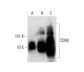
Date published: 2025-9-6

021-6093-6350

SCBT Portrait Logo
Seach Input

CD68 抗体 (KP1): sc-20060

4.4(45)
寫評論提問

说明书
  • CD68 抗体 KP1 是小鼠单克隆 IgG1 κ,CD68抗体, 在249篇文献中引用,规格为200 µg/ml
  • 与human肺泡巨噬细胞的亚细胞部分杂交
  • CD68 抗体 (KP1) 推荐用于 WB, IP, IF, IHC(P) 和 FCM,检测mouse, rat 和human 来源的 CD68
  • 抗CD68抗体(KP1)可与琼脂糖结合用于IP;与HRP结合用于WB、IHC(P)和ELISA;与藻红蛋白FITC结合用于IF、IHC(P)和FCM
  • 还可偶联Alexa Fluor® 488, Alexa Fluor® 546, Alexa Fluor® 594Alexa Fluor® 647,用于WB (RGB), IF, IHC(P) 和 FCM, 以及用于RGB荧光成像系统,例如iBright™ FL1000, FluorChem™, Typhoon, Azure和其他类似的系统
  • 还可偶联Alexa Fluor® 680Alexa Fluor® 790, 用于WB (NIR), IF 和 FCM; 以及用于近红外(NIR)检测系统,如LI-COR®/Odyssey®, iBright™ FL1000, FluorChem™, Typhoon, Azure和类似系统
  • 还可偶联PerCP 以及 Alexa Fluor® 405 用于IF, IHC(P) and FCM
  • 关于如何获取CD68 (KP1): sc-20060免费10 µg小样,联系我们技术服务部门 (或者您当地的代理商)了解详情。
  • m-IgG Fc BP-HRP, m-IgG1 BP-HRPm-IgGκ BP-HRP 是 CD68 抗体 (KP1) 在 WB and IHC(P) 应用中的首选二抗检测试剂。这些试剂现与CD68 抗体 (KP1) 组合成套装提供。 (请参阅下面的订购信息)。

快捷链接

関連項目

CD68抗体(KP1)是一种小鼠单克隆IgG1 kappa轻链抗体,可通过蛋白质印迹(WB)、免疫沉淀(IP)、免疫荧光(IF)、免疫组织化学和流式细胞术(FCM)检测小鼠、大鼠和人源CD68蛋白。CD68 (KP1) 抗体有非结合型和多种结合型,包括琼脂糖、辣根过氧化物酶 (HRP)、藻红蛋白 (PE)、异硫氰酸荧光素 (FITC) 和多种 Alexa Fluor® 结合物。CD68蛋白与鼠抗原巨细胞素同源,属于酸性、高度糖基化的溶酶体糖蛋白家族,包括lamp-1和lamp-2。CD68(KP1)单克隆抗体可识别一种在免疫系统中发挥关键作用的蛋白质,主要作为巨噬细胞和其他对吞噬作用和细胞碎片清除至关重要的髓细胞标记物。CD68蛋白主要存在于细胞质颗粒和各种非造血组织的细胞质中,例如肝、肾小管和肾小球,并出现在巨噬细胞、单核细胞、中性粒细胞、嗜碱性粒细胞和大型淋巴细胞的表面。这些免疫细胞上的CD68对于激活和功能至关重要,有助于识别病原体和摄取凋亡细胞,从而促进人体的防御机制和组织稳态。

仅限研究使用。不适用于诊断和治疗用途。

Alexa Fluor® 是Molecular Probes Inc., OR., USA的商标

LI-COR®和 Odyssey® 是LI-COR Biosciences的注册商标

CD68 抗体 (KP1) 参考文献:

  1. 溶酶体膜糖蛋白。结构, 生物合成和细胞内运输。  |  Fukuda, M. 1991. J Biol Chem. 266: 21327-30. PMID: 1939168
  2. CD68 巨噬细胞/骨髓相关抗原的分布。  |  Pulford, KA., et al. 1990. Int Immunol. 2: 973-80. PMID: 2078523
  3. 大鼠附睾中CD68糖蛋白的表达。  |  Liguori, G., et al. 2015. Biochimie. 118: 221-4. PMID: 26433032
  4. 妊娠糖尿病母亲胎盘中的CD68表达:病例对照研究。  |  Gosain, R., et al. 2023. Indian J Pathol Microbiol. 66: 727-731. PMID: 38084523
  5. 斑块内新生血管、CD68+和iNOS2+巨噬细胞浸润强度与严重颈动脉粥样硬化的粥样血栓形成和斑块内出血有关。  |  Balmos, IA., et al. 2023. Biomedicines. 11: PMID: 38137496
  6. 揭示CD8、CD68和VISTA作为胰腺导管腺癌患者诊断和预后标志物的潜力。  |  Rezagholizadeh, F., et al. 2024. Front Immunol. 15: 1283364. PMID: 38357542
  7. 肿瘤微环境中 CD68+SHP2+ 巨噬细胞的空间相互作用和功能状态与 NSCLC 的总生存率相关。  |  Liu, X., et al. 2024. Front Immunol. 15: 1396719. PMID: 38799432
  8. SPI1+CD68+巨噬细胞作为胃癌转移的生物标志物:联合抗血管生成和免疫治疗策略的理论依据。  |  Deng, G., et al. 2024. J Immunother Cancer. 12: PMID: 39455096
  9. 94-97-kDa的小鼠巨噬细胞膜蛋白能识别氧化的低密度脂蛋白和富含磷脂酰丝氨酸的脂质体,它与人类CD68的小鼠同源物--巨噬细胞蛋白相同。  |  Ramprasad, MP., et al. 1995. Proc Natl Acad Sci U S A. 92: 9580-4. PMID: 7568176
  10. 正常和恶性造血过程中细胞内 CD68 分子表达的流式细胞分析。  |  Strobl, H., et al. 1995. Br J Haematol. 90: 774-82. PMID: 7669655
  11. 与溶酶体糖蛋白有关的人类巨噬细胞标记物 CD68 的分子克隆。  |  Holness, CL. and Simmons, DL. 1993. Blood. 81: 1607-13. PMID: 7680921
  12. 小鼠大鼠蛋白和人类 CD68 的细胞表面表达及其作为氧化低密度脂蛋白巨噬细胞受体的作用。  |  Ramprasad, MP., et al. 1996. Proc Natl Acad Sci U S A. 93: 14833-8. PMID: 8962141

订购信息

产品名称产品编号规格价格数量收藏夹

CD68 抗体 (KP1)

sc-20060
200 µg/ml
$316.00

CD68 (KP1): m-IgG Fc BP-HRP 套装

sc-528358
200 µg Ab; 10 µg BP
$354.00

CD68 (KP1): m-IgGκ BP-HRP 套装

sc-520717
200 µg Ab, 40 µg BP
$354.00

CD68 (KP1): m-IgG1 BP-HRP 套装

sc-542889
200 µg Ab; 20 µg BP
$354.00

CD68 抗体 (KP1) AC

sc-20060 AC
500 µg/ml, 25% agarose
$416.00

CD68 抗体 (KP1) HRP

sc-20060 HRP
200 µg/ml
$316.00

CD68 抗体 (KP1) FITC

sc-20060 FITC
200 µg/ml
$330.00

CD68 抗体 (KP1) PE

sc-20060 PE
200 µg/ml
$343.00

CD68 抗体 (KP1) Alexa Fluor® 488

sc-20060 AF488
200 µg/ml
$357.00

CD68 抗体 (KP1) Alexa Fluor® 546

sc-20060 AF546
200 µg/ml
$357.00

CD68 抗体 (KP1) Alexa Fluor® 594

sc-20060 AF594
200 µg/ml
$357.00

CD68 抗体 (KP1) Alexa Fluor® 647

sc-20060 AF647
200 µg/ml
$357.00

CD68 抗体 (KP1) Alexa Fluor® 680

sc-20060 AF680
200 µg/ml
$357.00

CD68 抗体 (KP1) Alexa Fluor® 790

sc-20060 AF790
200 µg/ml
$357.00

CD68 抗体 (KP1) Alexa Fluor® 405

sc-20060 AF405
200 µg/ml
$357.00

CD68 抗体 (KP1) PerCP

sc-20060 PerCP
100 tests in 2 ml
$351.00

Hello, Would the the CD68 antibody (sc-20060) also work on porcine macrophages like it does on humans, mice, and rats for IHC-P? Best wishes, Tamara

Asked by: Tamara
Thank you for your question. There is a 65% homology in porcine. We only recommend a specie if the homology is 95%or higher .
Answered by: BlakeJ
Date published: 2024-05-28

Would this antibody work in canine tissue?

Asked by: maggie22
Thank you for your question . We would not recommend this antibody for canine. There is a 62% homology in canine .We only warranty a specie if the homology is 95% or higher.
Answered by: BlakeJ
Date published: 2023-06-07

What antigen retrieval method would you suggest for IHC-P?

Asked by: livlab
Thank you for your question. You can find our recommended method for antigen retrieval in our standart protocol, which is available under this link: https://www.scbt.com/resources/protocols/immunoperoxidase-staining
Answered by: Technical Support Europe
Date published: 2020-10-21

Were the THP-1 in the IF staining photos differentiated?

Asked by: ttam
Thank you for your question. No, this is a standard whole cell lysate that was used for this IF staining. The status of proteins in a cell lysate can vary, so localization of proteins can vary if depending on the status of the protein in the cells. We did not treat the cells in any special way.
Answered by: Tech Service
Date published: 2019-10-11

Hello! I am working on IHC-P project involving pigs heart tissue. would this antibody be suitable to work on pig tissues formalin fixed / paraffin embedded? Thanks

Asked by: Drzaid
Thank you for your question. We are not aware of any data or product citations using this antibody in pig samples.
Answered by: Technical Support
Date published: 2022-11-11

What kind of optimalisation (citrate/EDTA) does the antibody need for IHC-P on human brain tissue. I'd like to label microglia. Thanks

Asked by: doradavid11
Thank you for your question. We recommend using sodium citrate buffer (pH 6) with heat treatment for antigen retrieval with this antibody on FFPE. Please see our protocol for more details.
Answered by: Technical Support
Date published: 2023-12-29

When I use this antibody to run a flow cytometry experiment, should I apply a intracellular stain protocol or stain it in a surface staining way

Asked by: rhone
Thank you for your question. We recommend using a direct staining FCM protocol when using this mouse monoclonal antibody, CD68 (KP1): sc-20060. A link to our recommended protocol is provided below. https://www.scbt.com/scbt/resources/protocols/flow-cytometry
Answered by: Technical Support
Date published: 2023-12-29

What region of the protein does this antibody bind to? I see that it is reactive towards several species, so is it a conserved sequence?

Asked by: treedee21
Thank you for your question. CD68 Antibody (KP1) is a mouse monoclonal raised against a subcellular fraction of human alveolar macrophages. The exact epitope recognized by this antibody has not been determined.
Answered by: Technical Support
Date published: 2023-12-29
  • y_2025, m_8, d_27, h_5CST
  • bvseo_bulk, prod_bvqa, vn_bulk_3.0.42
  • cp_1, bvpage1
  • co_hasquestionsanswers, tq_10
  • loc_zh_CN, sid_20060, prod, sort_[SortEntry(order=LAST_APPROVED_ANSWER_SUBMISSION_TIME, direction=DESCENDING)]
  • clientName_scbt
  • bvseo_sdk, java_sdk, bvseo-4.0.0
  • CLOUD, getContent, 100ms
  • QUESTIONS, PRODUCT
Rated 5 out of 5 by from CD68-AF647Bought conjugated CD68-AF647. Not very good. Tried with IHC and IF for FFPE tissue and THP-1 cells. IF for FFPE did not work. IHC FFPE works reasonably well (this was a AF647 conjugated antibody, so this is not it's intended use). IF THP-1 very weak (1:50 - 1:100). I would recommend buying the unconjugated antibody and use with a secondary.
Date published: 2025-07-02
Rated 5 out of 5 by from works perfectI performed IHC staining using this antibody in human atherosclerosis sample and got strong and specific signal. I will buy it again.
Date published: 2023-09-24
Rated 5 out of 5 by from This antibody works very well for IHQThis antibody works very well for immunhistochemistry (mouse lung tissue)
Date published: 2023-06-20
Rated 4 out of 5 by from sc-20060It was good for IHC experiment with high specificity.
Date published: 2022-09-26
Rated 4 out of 5 by from Good signal in WB studiesIt works well in western blot studies using non-parenchymal cells (enriched in macrophages) isolated from rat livers
Date published: 2021-01-18
Rated 5 out of 5 by from Good performanceSeveral months ago, we purchased the CD68 anti-body, and the histochemical result was very good.
Date published: 2020-10-09
Rated 5 out of 5 by from A very nice antibody.The antibody has high sensitivity and strong specificity, as well as its accurate positioning and clear staining background.
Date published: 2020-05-15
Rated 5 out of 5 by from Good performanceOne year ago, we purchased the antibody against CD68 (sc-20060) . The antibody works well.
Date published: 2019-08-26
  • y_2025, m_8, d_27, h_5
  • bvseo_bulk, prod_bvrr, vn_bulk_3.0.42
  • cp_1, bvpage1
  • co_hasreviews, tv_0, tr_46
  • loc_zh_CN, sid_20060, prod, sort_[SortEntry(order=SUBMISSION_TIME, direction=DESCENDING)]
  • clientName_scbt
  • bvseo_sdk, java_sdk, bvseo-4.0.0
  • CLOUD, getReviews, 14ms
  • REVIEWS, PRODUCT
CD68 抗体 (KP1) is rated 4.4 out of 5 by 46.
  • y_2025, m_8, d_27, h_5
  • bvseo_bulk, prod_bvrr, vn_bulk_3.0.42
  • cp_1, bvpage1
  • co_hasreviews, tv_0, tr_46
  • loc_zh_CN, sid_20060, prod, sort_[SortEntry(order=SUBMISSION_TIME, direction=DESCENDING)]
  • clientName_scbt
  • bvseo_sdk, java_sdk, bvseo-4.0.0
  • CLOUD, getAggregateRating, 108ms
  • REVIEWS, PRODUCT