Date published: 2026-5-21

1-800-457-3801

SCBT Portrait Logo
Seach Input

Src Antibody (17AT28): sc-130124

4.6(12)
Write a reviewAsk a question

Datasheets
  • Src Antibody (17AT28) is a mouse monoclonal IgG1, cited in 33 publications, provided at 100 µg/ml
  • raised against full-length recombinant c-Src of human origin
  • recommended for detection of c-Src of mouse, rat and human origin by WB, IP, IF, IHC(P) and ELISA
  • See Src (B-12): sc-8056 for Src antibody conjugates, including AC, HRP, FITC, PE, Alexa Fluor® 488, 594, 647, 680 and 790.
  • At present, we have not yet completed the identification of the preferred secondary detection reagent(s) for Src Antibody (17AT28). This work is in progress.
Gene Editing Promo Banner

QUICK LINKS

SEE ALSO...

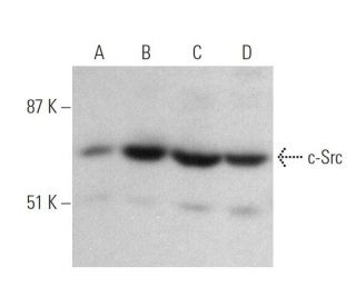
c-Src Antibody (17AT28) is a mouse monoclonal antibody raised against full-length recombinant human c-Src, a crucial member of the Src family kinases involved in cellular signal transduction. c-Src mouse monoclonal antibody (17AT28) recognizes an epitope within the c-Src immunogen sequence and demonstrates broad species cross-reactivity, detecting Src family kinases in mouse, rat, and human samples through Western blotting (WB), immunoprecipitation (IP), immunofluorescence (IF), immunohistochemistry on paraffin-embedded sections (IHCP), and enzyme-linked immunosorbent assay (ELISA) applications. The Src family comprises nine members in vertebrates, including c-Src, c-Yes, Fgr, Yrk, Fyn, Lyn, Hck, Lck, and Blk. These kinases share conserved structural features: an amino-terminal membrane anchor followed by SH3 and SH2 domains, which facilitate protein-protein interactions and signal transduction. c-Src, also known as pp60Src or proto-oncogene tyrosine-protein kinase Src, undergoes post-translational lipid modifications that anchor c-Src to the inner surface of the plasma membrane. This specific subcellular localization positions c-Src optimally for interaction with cell surface receptors and adhesion molecules, enabling c-Src to transmit signals that regulate cellular processes including proliferation, differentiation, motility, and adhesion. When c-Src becomes misregulated or aberrantly activated, the resulting dysregulation can promote uncontrolled cell growth and contribute to cancer progression. Anti-c-Src monoclonal antibody (17AT28) serves as an invaluable tool for researchers investigating signal transduction pathways, cellular growth regulation, and oncogenic processes.

For Research Use Only. Not Intended for Diagnostic or Therapeutic Use.

Ordering Information

Product NameCatalog #UNITPriceQtyFAVORITES

Src Antibody (17AT28)

sc-130124
100 µg/ml
$322.00

Can sc-130124: c-Src (17AT28) monoclonal antibody be used to stain formalin-fixed, paraffin-embedded (FFPE) tissue sections?

Asked by: Professor Griffin
Thank you for your question. Yes, sc-130124: c-Src (17AT28) is recommended for use in IHC with paraffin-embedded sections. We recommend performing antigen retrieval with sodium citrate buffer (pH 6) and heat. The full protocol can be found here: https://www.scbt.com/scbt/resources/protocols/immunoperoxidase-staining
Answered by: Technical Support
Date published: 2017-03-24
  • y_2026, m_5, d_20, h_12CST
  • bvseo_bulk, prod_bvqa, vn_bulk_3.0.46
  • cp_1, bvpage1
  • co_hasquestionsanswers, tq_1
  • loc_en_US, sid_130124, prod, sort_[SortEntry(order=LAST_APPROVED_ANSWER_SUBMISSION_TIME, direction=DESCENDING)]
  • clientName_scbt
  • bvseo_sdk, java_sdk, bvseo-4.0.0
  • CLOUD, getContent, 79ms
  • QUESTIONS, PRODUCT
Rated 4 out of 5 by from It can be usedMy protein samples were from RAW 264.7 cells, and I got a shallow band.
Date published: 2018-05-11
Rated 5 out of 5 by from Works great!Yielded nice strong band when used at 1:200 for mouse tissue lysates.
Date published: 2018-03-23
Rated 5 out of 5 by from 非常棒!很清楚跑的很清楚!照片暂时不能放!因为文章还没出来!买这个是一个明智的选择!!!!!!!!!!!!!!!!!
Date published: 2018-02-02
Rated 5 out of 5 by from Excellent antibodyThis antibody works very well for western blot analysis.
Date published: 2017-11-14
Rated 4 out of 5 by from great feature, great productalways love it, give us consistent result, easy to use
Date published: 2017-07-24
Rated 5 out of 5 by from WB resutl is very goodWe have been using this antibody consistently in our research with mouse brain tissue for WB analysis and never meet problems.
Date published: 2017-05-13
Rated 5 out of 5 by from strong bandWe observed strong band in WB, dilution of primary antibody is 1: 300.
Date published: 2017-05-13
Rated 4 out of 5 by from Good antibody and good signals (primary canineGood antibody and good signals (primary canine chondrocytes)
Date published: 2016-01-29
  • y_2026, m_5, d_21, h_10
  • bvseo_bulk, prod_bvrr, vn_bulk_3.0.46
  • cp_1, bvpage1
  • co_hasreviews, tv_0, tr_12
  • loc_en_US, sid_130124, prod, sort_[SortEntry(order=SUBMISSION_TIME, direction=DESCENDING)]
  • clientName_scbt
  • bvseo_sdk, java_sdk, bvseo-4.0.0
  • CLOUD, getReviews, 11ms
  • REVIEWS, PRODUCT
Src Antibody (17AT28) is rated 4.6 out of 5 by 12.
  • y_2026, m_5, d_21, h_10
  • bvseo_bulk, prod_bvrr, vn_bulk_3.0.46
  • cp_1, bvpage1
  • co_hasreviews, tv_0, tr_12
  • loc_en_US, sid_130124, prod, sort_[SortEntry(order=SUBMISSION_TIME, direction=DESCENDING)]
  • clientName_scbt
  • bvseo_sdk, java_sdk, bvseo-4.0.0
  • CLOUD, getAggregateRating, 99ms
  • REVIEWS, PRODUCT