")
PKC alpha Antibody (H-7): sc-8393
- PKC alpha Antibody (H-7) is a mouse monoclonal IgG1 κ PKC alpha antibody, cited in 324 publications, provided at 200 µg/ml
- specific for an epitope mapping between amino acids 651-672 at the C-terminus of PKC α of human origin
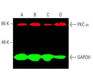
- PKC alpha Antibody (H-7) is recommended for detection of PKC α of mouse, rat and human origin by WB, IP, IF, IHC(P) and ELISA
- Anti-PKC alpha Antibody (H-7) is available conjugated to agarose for IP; HRP for WB, IHC(P) and ELISA; and to either phycoerythrin or FITC for IF, IHC(P) and FCM
- also available conjugated to Alexa Fluor® 488, Alexa Fluor® 546, Alexa Fluor® 594 or Alexa Fluor® 647 for WB (RGB), IF, IHC(P) and FCM, and for use with RGB fluorescent imaging systems, such as iBright™ FL1000, FluorChem™, Typhoon, Azure and other comparable systems
- also available conjugated to Alexa Fluor® 680 or Alexa Fluor® 790 for WB (NIR), IF and FCM; for use with Near-Infrared (NIR) detection systems, such as LI-COR®Odyssey®, iBright™ FL1000, FluorChem™, Typhoon, Azure and other comparable systems
- m-IgG Fc BP-HRP, m-IgG1 BP-HRP and m-IgGκ BP-HRP are the preferred secondary detection reagents for PKC alpha Antibody (H-7) for WB and IHC(P) applications. These reagents are now offered in bundles with PKC alpha Antibody (H-7) (see ordering information below).

PKC α Antibody (H-7) is a mouse monoclonal IgG1 kappa light chain antibody that detects PKC alpha protein of mouse, rat, and human origin by western blotting (WB), immunoprecipitation (IP), immunofluorescence (IF), immunohistochemistry, and enzyme-linked immunosorbent assay (ELISA). PKC α Antibody (H-7) is available in both non-conjugated and various conjugated forms, including agarose, horseradish peroxidase (HRP), phycoerythrin (PE), fluorescein isothiocyanate (FITC), and multiple Alexa Fluor® conjugates. PKC alpha protein, a member of the protein kinase C (PKC) family, plays a crucial role in regulating numerous cellular functions such as cell growth, differentiation, gene expression, and hormone secretion, making PKC alpha vital for maintaining cellular homeostasis. PKC alpha is primarily located in the cytoplasm and translocates to the cell membrane upon activation, which is essential for PKC alpha′s role in signal transduction pathways. This translocation is triggered by diacylglycerols (DAG) and tumor-promoting phorbol esters, which bind to and activate PKC alpha, allowing PKC alpha to phosphorylate target proteins and modulate various cellular responses. The diverse expression patterns of PKC isoforms across different tissues highlight PKC alpha′s importance in specific signaling pathways, and PKC alpha′s unique cofactor dependencies further underscore PKC alpha′s distinct regulatory mechanisms within the PKC family.
Alexa Fluor® is a trademark of Molecular Probes Inc., OR., USA
LI-COR® and Odyssey® are registered trademarks of LI-COR Biosciences
PKC alpha Antibody (H-7) References:
- The role of protein kinase C-alpha (PKC-alpha) in melanoma. | Lahn, MM. and Sundell, KL. 2004. Melanoma Res. 14: 85-9. PMID: 15057036
- A new member of the protein kinase C family, nPKC theta, predominantly expressed in skeletal muscle. | Osada, S., et al. 1992. Mol Cell Biol. 12: 3930-8. PMID: 1508194
- Resveratrol regulates cellular PKC alpha and delta to inhibit growth and induce apoptosis in gastric cancer cells. | Atten, MJ., et al. 2005. Invest New Drugs. 23: 111-9. PMID: 15744586
- Structural and functional diversities of a family of signal transducing protein kinases, protein kinase C family; two distinct classes of PKC, conventional cPKC and novel nPKC. | Ohno, S., et al. 1991. Adv Enzyme Regul. 31: 287-303. PMID: 1877391
- Expression and characterization of protein kinase C-delta. | Olivier, AR. and Parker, PJ. 1991. Eur J Biochem. 200: 805-10. PMID: 1915352
- PKC-alpha modulates TGF-beta signaling and impairs podocyte survival. | Tossidou, I., et al. 2009. Cell Physiol Biochem. 24: 627-34. PMID: 19910703
- Coronavirus disease 2019 (COVID-19), human erythrocytes and the PKC-alpha/-beta inhibitor chelerythrine -possible therapeutic implication. | Ghashghaeinia, M., et al. 2020. Cell Cycle. 19: 3399-3405. PMID: 33305655
- Calcium-dependent activation of a multifunctional protein kinase by membrane phospholipids. | Takai, Y., et al. 1979. J Biol Chem. 254: 3692-5. PMID: 438153
- Turnover of inositol phospholipids and signal transduction. | Nishizuka, Y. 1984. Science. 225: 1365-70. PMID: 6147898
- The role of protein kinase C in cell surface signal transduction and tumour promotion. | Nishizuka, Y. Nature. 308: 693-8. PMID: 6232463
- Protein kinase C as a possible receptor protein of tumor-promoting phorbol esters. | Kikkawa, U., et al. 1983. J Biol Chem. 258: 11442-5. PMID: 6311812
- Direct activation of calcium-activated, phospholipid-dependent protein kinase by tumor-promoting phorbol esters. | Castagna, M., et al. 1982. J Biol Chem. 257: 7847-51. PMID: 7085651
Ordering Information
| Product Name | Catalog # | UNIT | Price | Qty | FAVORITES | |
PKC alpha Antibody (H-7) | sc-8393 | 200 µg/ml | $322.00 | |||
PKC alpha Antibody (H-7): m-IgG Fc BP-HRP Bundle | sc-528219 | 200 µg Ab; 10 µg BP | $361.00 | |||
PKC alpha Antibody (H-7): m-IgGκ BP-HRP Bundle | sc-520544 | 200 µg Ab, 40 µg BP | $361.00 | |||
PKC alpha Antibody (H-7): m-IgG1 BP-HRP Bundle | sc-542822 | 200 µg Ab; 20 µg BP | $361.00 | |||
PKC alpha Antibody (H-7) AC | sc-8393 AC | 500 µg/ml, 25% agarose | $424.00 | |||
PKC alpha Antibody (H-7) HRP | sc-8393 HRP | 200 µg/ml | $322.00 | |||
PKC alpha Antibody (H-7) FITC | sc-8393 FITC | 200 µg/ml | $336.00 | |||
PKC alpha Antibody (H-7) PE | sc-8393 PE | 200 µg/ml | $349.00 | |||
PKC alpha Antibody (H-7) Alexa Fluor® 488 | sc-8393 AF488 | 200 µg/ml | $364.00 | |||
PKC alpha Antibody (H-7) Alexa Fluor® 546 | sc-8393 AF546 | 200 µg/ml | $364.00 | |||
PKC alpha Antibody (H-7) Alexa Fluor® 594 | sc-8393 AF594 | 200 µg/ml | $364.00 | |||
PKC alpha Antibody (H-7) Alexa Fluor® 647 | sc-8393 AF647 | 200 µg/ml | $364.00 | |||
PKC alpha Antibody (H-7) Alexa Fluor® 680 | sc-8393 AF680 | 200 µg/ml | $364.00 | |||
PKC alpha Antibody (H-7) Alexa Fluor® 790 | sc-8393 AF790 | 200 µg/ml | $364.00 | |||
PKC alpha (H-7) Neutralizing Peptide | sc-8393 P | 100 µg/0.5 ml | $69.00 |