")
Anticorps PELP1 (E-1): sc-390599
- L'anticorps PELP1 E-1 est un monoclonal IgM (chaîne légère kappa) L'Anticorps PELP1 fourni en 200 µg/ml
- spécifique d'une correspondance épitopique entre les acides aminés 37-62 de PELP1 de human d'origine
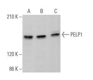
- recommandé pour la détection de PELP1 d'origine mouse, rat et human par WB, IP, IF et ELISA; également réactif avec d'autres espèces, dont bovine
- Le m-IgGκ BP-HRP est le réactif de détection secondaire préféré pour l'anticorps PELP1 (E-1) pour les applications WB. Ce réactif est désormais proposé en association avec l'anticorps PELP1 (E-1)(voir les informations relatives à la commande ci-dessous). Pour d'autres conjugués m-IgGκ BP, voir notre liste complète de protéines de liaison à l'IgG de souris.

ACCÈS RAPIDE AUX LIENS
VOIR ÉGALEMENT...
PELP1 Antibody (E-1) is an IgM κ mouse monoclonal PELP1 antibody (also designated PELP1 antibody) that detects the PELP1 protein of mouse, rat and human origin by WB, IP, IF and ELISA. PELP1 Antibody (E-1) is available as the non-conjugated anti-PELP1 antibody. The estrogen receptor (ER) plays an important role in cancer progression. PELP1/MNAR (proline-, glutamic acid- and leucine-rich protein-1/modulator of non-genomic activity of estrogen receptor), a novel coregulatory protein, modulates genomic as well as nongenomic activity of estrogen receptors. PELP1 plays an essential role in the proliferation of cancerous endometrial cells. PELP1 expression, in both the stroma and epithelial cells, and localization are widely deregulated in endometrial cancers. In addition, PELP1 and ER beta localize predominantly in the cytoplasm of high-grade endometrial tumors. PELP1 coactivates ER-mediated transcription and also serves as a corepressor of other nuclear hormone receptors (NR)- and non-NR sequence-specific transcription factors, including GR, Nur77, AP1, NFkappaB, and TCF/SRF. PELP1 participates in chromatin remodeling activity via displacement of histone 1 in cancer cells and is expressed in all stages of endometrium.
Alexa Fluor® est une marque déposée de Molecular Probes Inc., OR., USA
LI-COR® et Odyssey® sont marques déposées de LI-COR Biosciences
Anticorps PELP1 (E-1) Références:
- Interactions fonctionnelles entre le coactivateur du récepteur des œstrogènes PELP1/MNAR et la protéine du rétinoblastome. | Balasenthil, S. and Vadlamudi, RK. 2003. J Biol Chem. 278: 22119-27. PMID: 12682072
- Clonage et caractérisation fonctionnelle du promoteur de PELP1/MNAR. | Mishra, SK., et al. 2004. Gene. 330: 115-22. PMID: 15087130
- Rôle potentiel d'un nouveau coactivateur transcriptionnel PELP1 dans le déplacement de l'histone H1 dans les cellules cancéreuses. | Nair, SS., et al. 2004. Cancer Res. 64: 6416-23. PMID: 15374949
- Le corépresseur transcriptionnel PELP1 recrute HDAC2 et masque les histones à l'aide de deux domaines distincts. | Choi, YB., et al. 2004. J Biol Chem. 279: 50930-41. PMID: 15456770
- La suppression de PELP1/MNAR inhibe la prolifération et la métastase des cellules de carcinome endométrial. | Wan, J. and Li, X. 2012. Oncol Rep. 28: 2035-42. PMID: 22992812
Informations pour la commande
| Nom du produit | Ref. Catalogue | COND. | Prix HT | QTÉ | Favoris | |
Anticorps PELP1 (E-1) | sc-390599 | 200 µg/ml | $322.00 | |||
PELP1 (E-1): m-IgGκ BP-HRP Kit | sc-552530 | 200 µg Ab; 40 µg BP | $361.00 | |||
PELP1 (E-1) peptide neutralisant | sc-390599 P | 100 µg/0.5 ml | $69.00 |